SECTION 5.1
Arteries, veins, capillaries and anastomoses
Compare arteries, veins and capillaries by wall structure and function, and explain what an anastomosis provides.
Learn
Blood vessels form a closed circuit with three main kinds of tube. Arteries carry blood away from the heart under high pressure. Veins carry blood back to the heart at low pressure. Between them, capillaries form the exchange surface where substances move between blood and tissue. All three have the same basic three-layer wall, but the proportions differ, and those differences explain their behaviour. Arteries have a thick middle layer of muscle and elastic tissue to withstand and regulate pressure. Capillaries are reduced to a single endothelial layer so that exchange can happen quickly. Veins have thin walls and a wide lumen, and they contain valves to keep blood moving in one direction. Where two vessels join, they form an anastomosis, which can provide an alternative route if one is blocked. This section compares the three types and introduces that safety feature.
Key Concepts & Topics
Three tunics, three vessel types
Most blood vessels have three layers, called tunics. The tunica intima is the innermost layer, made of a smooth endothelial lining that reduces friction with flowing blood. The tunica media is the middle layer, containing smooth muscle and elastic fibres; it controls the vessel’s diameter and helps withstand pressure. The tunica externa is the outer layer of connective tissue that anchors the vessel to surrounding structures. The lumen is the hollow space through which blood flows. Vessels differ mainly in the relative thickness of these layers: arteries have a thick tunica media, capillaries have essentially only the intima, and veins have a thinner media but a larger lumen. Because muscle controls diameter, the arteries and arterioles are also the main site where the body adjusts blood flow to different organs.
For example: The smooth lining of the tunica intima is why normal blood does not clot inside a healthy vessel: a smooth surface does not activate platelets.
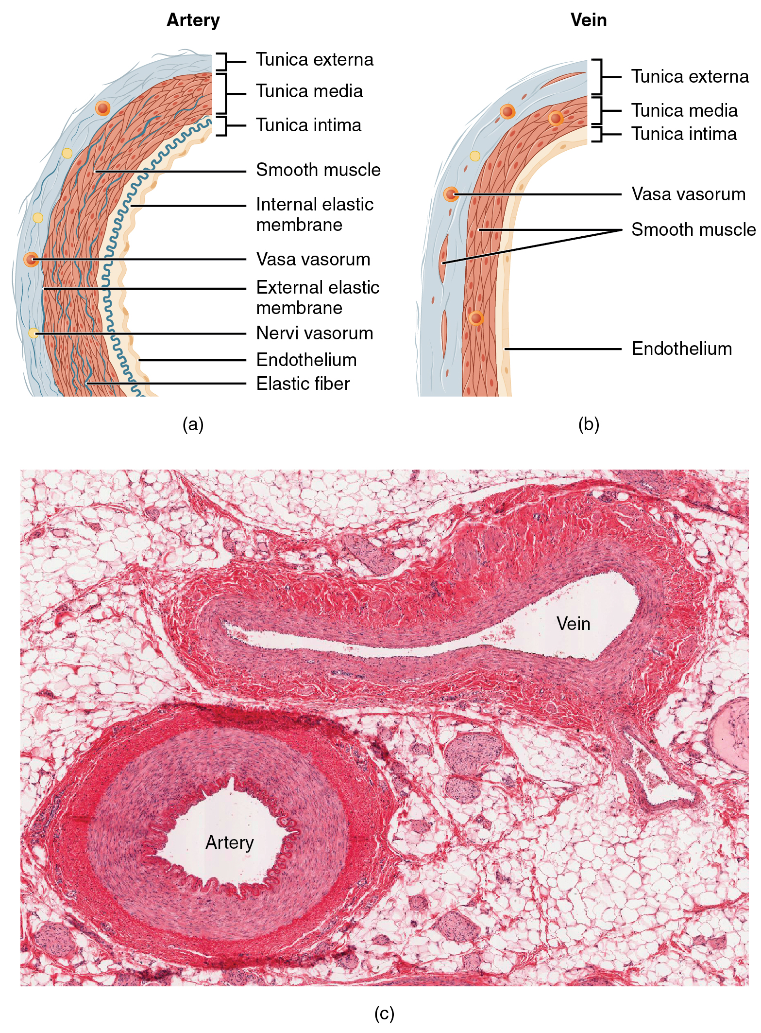 Enlarge illustration ↗
Start at the lumen—the open space carrying blood—and work outward through the layers. Compare the muscular middle layer. A vessel is a living, layered structure, not an empty rigid pipe.
Image source and licence
OpenStax College · Source image · CC BY 3.0. Original image, reproduced without alteration.
Arteries: pressure and control
Arteries carry blood away from the heart and are built to handle the pressure the heart generates. Their thick tunica media contains both elastic fibres and smooth muscle. Elastic arteries, such as the aorta, stretch as the heart ejects blood and recoil between beats, which smooths the pressure pulse and keeps blood moving during diastole. Muscular arteries distribute blood to organs and can contract or relax to change their diameter, a process called vasoconstriction or vasodilation. The smallest arteries are arterioles, which lead into capillary networks and are the main control point for local blood flow. Constricting an arteriole reduces flow to that tissue; dilating it increases flow. This control lets the body send more blood to working muscles and less to resting organs without changing total output.
For example: During exercise, arterioles in working muscle dilate while those in the gut constrict, so blood is redirected without the heart having to supply every tissue at once.
Capillaries: the exchange surface
Capillaries are the smallest vessels and the only place where exchange between blood and tissue occurs at a useful rate. Their wall is essentially a single layer of endothelial cells resting on a basement membrane, so the distance substances must cross is extremely short. Three structural types suit different needs. Continuous capillaries have tightly joined endothelial cells and are found in skin, muscle and lungs; they allow small molecules and water through but restrict larger ones. Fenestrated capillaries have pores that speed the movement of fluid and small solutes, and are found in the kidney, intestine and endocrine glands. Sinusoids are wider, leakier channels found in the liver, spleen and bone marrow, allowing proteins and even cells to pass. Blood flow through a capillary bed can also be adjusted by precapillary sphincters, which open and close to direct flow where it is needed.
For example: The air sacs of the lungs are wrapped in continuous capillaries so thin that oxygen has only a short distance to travel into the blood.
Veins: volume and return
Veins carry blood back to the heart and are adapted to low pressure. Their walls are thinner than those of arteries, with less muscle and elastic tissue, and their lumen is wider, so they offer little resistance to flow. They hold a large share of the body’s blood volume at any moment, which is why they are called capacitance vessels. Blood in the limbs must travel upwards against gravity, so many veins contain valves, folds of the lining that allow flow towards the heart but prevent it from falling back. Movement of the limbs helps squeeze the veins, and this skeletal muscle pump, together with breathing movements, assists return. If valves weaken, blood can pool and the vein becomes enlarged and twisted, which is a varicose vein.
For example: Standing still for a long time allows blood to pool in the leg veins, which is why walking and contracting the calf muscles help prevent swelling and clots.
Anastomoses and collateral circulation
An anastomosis is a connection between two blood vessels that normally supply the same region. It provides an alternative route: if one vessel is blocked, blood can still reach the tissue through the other. This is collateral circulation, and it is well developed in the joints, the stomach, the intestines and parts of the limbs and brain. Not every vessel has this safety net. An end artery, also called a terminal artery, is the only supply to a region, with no effective anastomosis; if it blocks, the tissue it supplies is at risk of dying. The coronary arteries, the renal arteries and parts of the brain behave in this way, which is why a blockage there can cause infarction. The presence or absence of anastomoses therefore helps predict what a blockage will do.
For example: The palm and the joints have rich anastomoses, so a single blocked artery often does not cause tissue death there; the heart has few, so a coronary blockage is dangerous.
Key facts to remember
- Most vessels have three tunics: intima (endothelium), media (muscle and elastic fibres) and externa (connective tissue).
- Arteries carry blood away from the heart, have a thick media and control flow through vasoconstriction and vasodilation of arterioles.
- Capillaries are a single endothelial layer; continuous, fenestrated and sinusoid types suit different exchange needs.
- Veins have thin walls, a wide lumen and valves, act as capacitance vessels and depend on the skeletal muscle pump for return.
- An anastomosis provides an alternative route; an end artery has no effective alternative, so its blockage threatens the tissue.
Example · Compare three tubes in one circuit. The aorta is an elastic artery: its thick wall stretches with each beat and recoils to keep blood moving. It branches into muscular arteries and then arterioles, whose diameter sets how much blood reaches each tissue. Between the arterioles and venules lie capillaries, one cell thick, where oxygen and nutrients enter the tissue and waste leaves. The venules and veins collect the blood; their wide, thin-walled tubes hold much of the blood volume, and their valves and the calf muscle pump push it back to the heart. Where two arteries join, as in the palm, an anastomosis means a blockage may not be fatal to the tissue; where no such join exists, as in the heart, it may be.
Read full notes
Read one topic at a time. These notes explain the mechanisms and relationships behind the key facts.
Three tunics, three vessel types
The three-layer wall is a common plan, and the differences between vessels are differences of proportion. The intima is always present, because every vessel needs a smooth, non-thrombogenic lining. The media varies enormously: thick and muscular in arteries, thin in veins, absent in capillaries. The externa anchors the vessel and merges with surrounding connective tissue. When you look at a vessel under the microscope, the first thing to notice is the thickness of the media relative to the lumen. A thick media with a relatively small lumen identifies an artery; a thin media with a wide lumen identifies a vein; a wall of essentially one cell layer identifies a capillary. Reading the proportions is more useful than memorising absolute sizes, because vessel size varies with the organ and with the person.
Arteries: pressure and control
Arteries solve two problems at once. They must withstand the pressure of each heartbeat without bursting, and they must allow the body to redirect blood where it is needed. Elastic tissue in the wall solves the first problem: it stretches during ejection and recoils afterwards, converting the intermittent push of the heart into a smoother flow. Smooth muscle solves the second: by contracting or relaxing, it changes the diameter of the vessel and therefore the resistance to flow. Because resistance depends strongly on diameter, small changes in an arteriole have large effects on flow. This is why the arterioles, rather than the large arteries, are described as the main resistance vessels and the main control point for distributing cardiac output.
Capillaries: the exchange surface
The capillary is where the circulatory system does its real work. Its wall is one cell thick, so diffusion distances are minimal, and its total surface area in the body is enormous. Blood flows slowly through capillary beds, which gives time for exchange. Two features vary to suit different tissues. Permeability determines what can cross: continuous capillaries restrict large molecules, fenestrated capillaries allow rapid fluid movement, and sinusoids allow proteins and cells through. Flow control determines where blood goes: precapillary sphincters open and close so that not every capillary in a bed is perfused at once. This is efficient, because perfusing all capillaries all the time would demand a much larger blood volume. It also explains why tissues can increase their capillary perfusion sharply when they become active.
Veins: volume and return
Veins are often treated as passive return pipes, but they perform two active roles. First, they store blood: because their walls are distensible, they hold a large fraction of the total blood volume and can release it when needed. Second, they help return blood against gravity. Valves prevent backflow, and the skeletal muscle pump uses the movement of limbs to push blood upwards; breathing also assists by changing pressure in the chest and abdomen. These mechanisms matter clinically. Prolonged immobility allows blood to pool and raises the risk of deep vein thrombosis, which is why early movement after surgery and compression stockings are used. Weakened valves cause varicose veins, and a failure of the muscle pump contributes to swelling in the legs.
Anastomoses and collateral circulation
Anastomoses show that the vascular system is a network, not a set of independent pipes. Where vessels join, a blockage in one can be compensated by flow through another, and the tissue may survive with reduced perfusion. The body can also enlarge existing anastomoses over time when a vessel narrows gradually, which is why some patients with slowly progressive coronary disease develop a collateral supply. The opposite situation is the end artery, where no alternative exists and a sudden blockage threatens the tissue immediately. Recognising which regions have good collateral supply, and which do not, helps explain why identical blockages produce very different outcomes and why the timing of a blockage matters as much as its location.
Explore the diagram
Start with the whole view, then select a numbered structure. Compare the additional illustrations to connect what a structure looks like with what it does.
Vessel walls match their jobs
Cross-sections at different enlargements. Compare wall-to-lumen proportions, not absolute diameters. The capillary wall is only one endothelial layer plus its basal support.
A thick smooth-muscle and elastic wall tolerates and modifies pulsatile pressure. Arteries carry blood away from the heart, regardless of oxygen content.
Connect the illustrations to the key ideas
ARTERY · thick media, high pressure
Elastic tissue smooths the pulse and smooth muscle controls diameter and flow.
CAPILLARY · one endothelial layer
Short diffusion distance and slow flow allow exchange; sphincters direct the flow.
VEIN AND ANASTOMOSIS · thin wall, wide lumen, valves; vessels join
Veins store volume and assist return; anastomoses provide an alternative route.
Core Concepts Defined
- Artery
- A vessel that carries blood away from the heart under relatively high pressure.
- Arteriole
- The smallest artery, leading into a capillary bed and controlling local blood flow.
- Vein
- A vessel that carries blood back to the heart at low pressure.
- Venule
- A small vessel that collects blood from capillaries and joins to form veins.
- Capillary
- A microscopic vessel with a single endothelial layer, where exchange between blood and tissue occurs.
- Tunica intima
- The innermost vessel layer, a smooth endothelial lining.
- Tunica media
- The middle layer of smooth muscle and elastic fibres that controls diameter.
- Tunica externa
- The outer connective-tissue layer that anchors the vessel.
- Lumen
- The hollow space inside a vessel through which blood flows.
- Endothelium
- The simple squamous lining of the inner vessel surface.
- Elastic artery
- A large artery with abundant elastic fibres that smooths the pressure pulse.
- Muscular artery
- A distributing artery with a thick muscular media that can change diameter.
- Vasoconstriction
- Narrowing of a vessel by contraction of its smooth muscle, reducing flow.
- Vasodilation
- Widening of a vessel by relaxation of its smooth muscle, increasing flow.
- Continuous capillary
- A capillary with tightly joined endothelial cells, found in skin, muscle and lungs.
- Fenestrated capillary
- A capillary with pores that speed fluid and solute movement, found in kidney and intestine.
- Sinusoid
- A wide, leaky capillary allowing proteins and cells to pass, found in liver, spleen and marrow.
- Valve (venous)
- A fold of vein lining that keeps blood flowing towards the heart.
- Capacitance vessel
- A vessel that holds a large share of blood volume; veins are the main example.
- Skeletal muscle pump
- The action of contracting limb muscles squeezing veins to assist blood return.
- Anastomosis
- A connection between two vessels supplying the same region, providing an alternative route.
- Collateral circulation
- Blood flow through an alternative route when the usual vessel is blocked.
- End (terminal) artery
- An artery that is the only supply to a region, with no effective anastomosis.
- Varicose vein
- An enlarged, twisted vein caused by weakened valves and pooled blood.
Go deeper · worked example
Why the same blockage has different effects
- Identify the tissue and its supply. A tissue may be supplied by a single end artery or by several arteries that join through anastomoses.
- Consider a blockage in a region with anastomoses. If one artery is blocked, blood can reach the tissue through the connecting vessel, so the tissue may survive with reduced flow.
- Consider a blockage in a region with an end artery. There is no alternative route, so the tissue beyond the blockage loses its supply and is at risk of infarction.
- Link to vessel structure. Arterioles can dilate to increase flow through the remaining route, but they cannot create a route that does not exist.
- Apply clinically. This is why a blocked artery in the hand or a joint is often less serious than a blocked coronary or renal artery, and why the presence of anastomoses is assessed in planning surgery.
Explain it yourself · Explain why the heart is more vulnerable to a blocked artery than the palm of the hand.
Reveal a model explanation
The palm has a rich anastomotic network, so if one artery is blocked, blood can reach the tissue through another route and the tissue may survive. The heart muscle is supplied by coronary arteries that behave largely as end arteries, with little effective alternative supply. When a coronary artery blocks, the muscle it supplies is deprived of oxygen and can infarct. The difference is not in the muscle but in the plumbing: the availability of a collateral route determines the consequence.
Check
Answer three questions from this section — one easy, one medium and one hard. If you miss one, you get a fresh set with explanations for the last attempt.
Practise this sectionOpen all questions and filter by difficulty or Bloom levelReferences and review status
OpenStax, Anatomy and Physiology 2e, 20.1 Structure and Function of Blood Vessels; Tortora & Derrickson, Principles of Anatomy and Physiology, 12e, ch. 21. Author review only; medical and French-language expert review pending.
Original teaching schematics. English and French wording is author-reviewed for study; anatomy and French-language expert review is pending. Checks are practice only, not a graded examination.
Practice progress is saved on this device for the current learner.