SECTION 8.1
Urinary organs and the nephron
Locate the urinary organs and identify the parts of the nephron, relating each part to its position in the kidney.
Learn
The urinary system removes waste products from the blood, regulates the volume and composition of body fluids, and produces urine. It consists of two kidneys, two ureters, the urinary bladder and the urethra. The kidneys do the real work: each contains about a million microscopic filters called nephrons, and each nephron has a renal corpuscle, where blood is filtered, and a renal tubule, where the filtrate is modified as it passes. The kidney itself is organised into an outer cortex and an inner medulla, and the tubules are arranged so that parts of the nephron lie in each region. This section locates the urinary organs, describes the internal structure of the kidney, and identifies the parts of the nephron and where each part sits.
Key Concepts & Topics
The urinary organs and their positions
The two kidneys are bean-shaped organs lying against the back wall of the abdomen on either side of the vertebral column, between about the twelfth thoracic and third lumbar vertebrae. The right kidney sits slightly lower than the left because the liver occupies space above it. Each kidney has a concave side facing inward, called the hilum, where the renal artery, renal vein and ureter enter or leave. From each hilum, a muscular tube called the ureter carries urine downward to the urinary bladder, a hollow muscular sac in the pelvis that stores urine. When it is emptied, urine leaves the body through the urethra. Because the bladder lies in the pelvis, it expands upward as it fills, which is why a full bladder can be felt above the pubic bone.
For example: Kidney pain is usually felt in the flank, the region of the back between the ribs and the hip, because that is where the kidneys lie.
Inside the kidney: cortex, medulla and pelvis
A kidney cut in half shows three regions. The outer cortex is a rim of tissue just beneath the capsule, and it contains the renal corpuscles of the nephrons. The medulla lies deep to the cortex and is divided into triangular structures called renal pyramids, whose tips point inward; the tubules that concentrate urine run through the pyramids. Between the pyramids, extensions of cortex called renal columns pass inward. The innermost space is the renal pelvis, a funnel-shaped cavity that collects urine from the pyramids through cup-like extensions called calyces and continues as the ureter. This arrangement is functional, not just descriptive: filtration happens in the cortex, and the concentration of urine depends on the loops and collecting ducts running through the medulla towards the pelvis.
For example: A kidney stone forming in the renal pelvis can block the flow of urine and cause severe flank pain that radiates toward the groin.
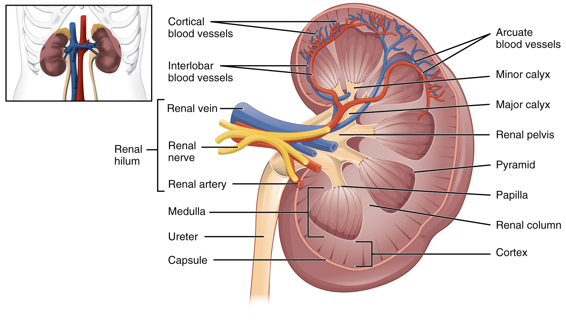 Enlarge illustration ↗
Trace urine from a pyramid tip into a calyx, then the renal pelvis and ureter. The cortex forms an outer band; the pyramids belong to the medulla. Blood vessels and urine-drainage spaces are different routes.
Image source and licence
OpenStax College · Source image · CC BY 3.0. Original image, reproduced without alteration.
The nephron: corpuscle and tubule
The nephron is the functional unit of the kidney, and each kidney contains about a million of them. Every nephron has two main parts. The renal corpuscle is a rounded structure in the cortex consisting of a tuft of capillaries called the glomerulus enclosed in a cup-shaped chamber called the glomerular (Bowman's) capsule. Blood enters the glomerulus through an afferent arteriole and leaves through an efferent arteriole, and fluid filtered from the blood collects in the capsule. The renal tubule begins at the capsule and continues as a coiled proximal convoluted tubule in the cortex, then a descending and ascending loop of Henle that dips into the medulla, then a distal convoluted tubule in the cortex again, and finally a collecting duct that passes through the medulla to empty into a calyx. The collecting duct receives several nephrons and is the last place where urine composition is adjusted.
For example: Because the loop of Henle dips into the medulla while the convoluted tubules stay in the cortex, a single nephron spans two kidney regions.
Common misconception: The collecting duct is not part of a single nephron. Several nephrons drain into one collecting duct, which carries urine to a calyx.
Blood supply of the nephron
The nephron has an unusual blood supply because it needs both high pressure for filtration and a second capillary bed for exchange. Blood reaches the kidney through the renal artery, which divides into progressively smaller vessels until an afferent arteriole enters each glomerulus. The glomerulus is a high-pressure capillary bed, and the pressure drives fluid out of the blood into the capsule. Blood then leaves through the efferent arteriole, which is narrower than the afferent vessel and therefore helps maintain that pressure. The efferent arteriole leads to a second capillary network, the peritubular capillaries, which wrap around the tubule, and to the vasa recta, long vessels that run alongside the loop of Henle into the medulla. These capillaries reclaim the water and solutes that the tubule reabsorbs and deliver substances the tubule secretes.
For example: Because the efferent arteriole is narrower than the afferent one, pressure stays high in the glomerulus, which is what makes filtration possible.
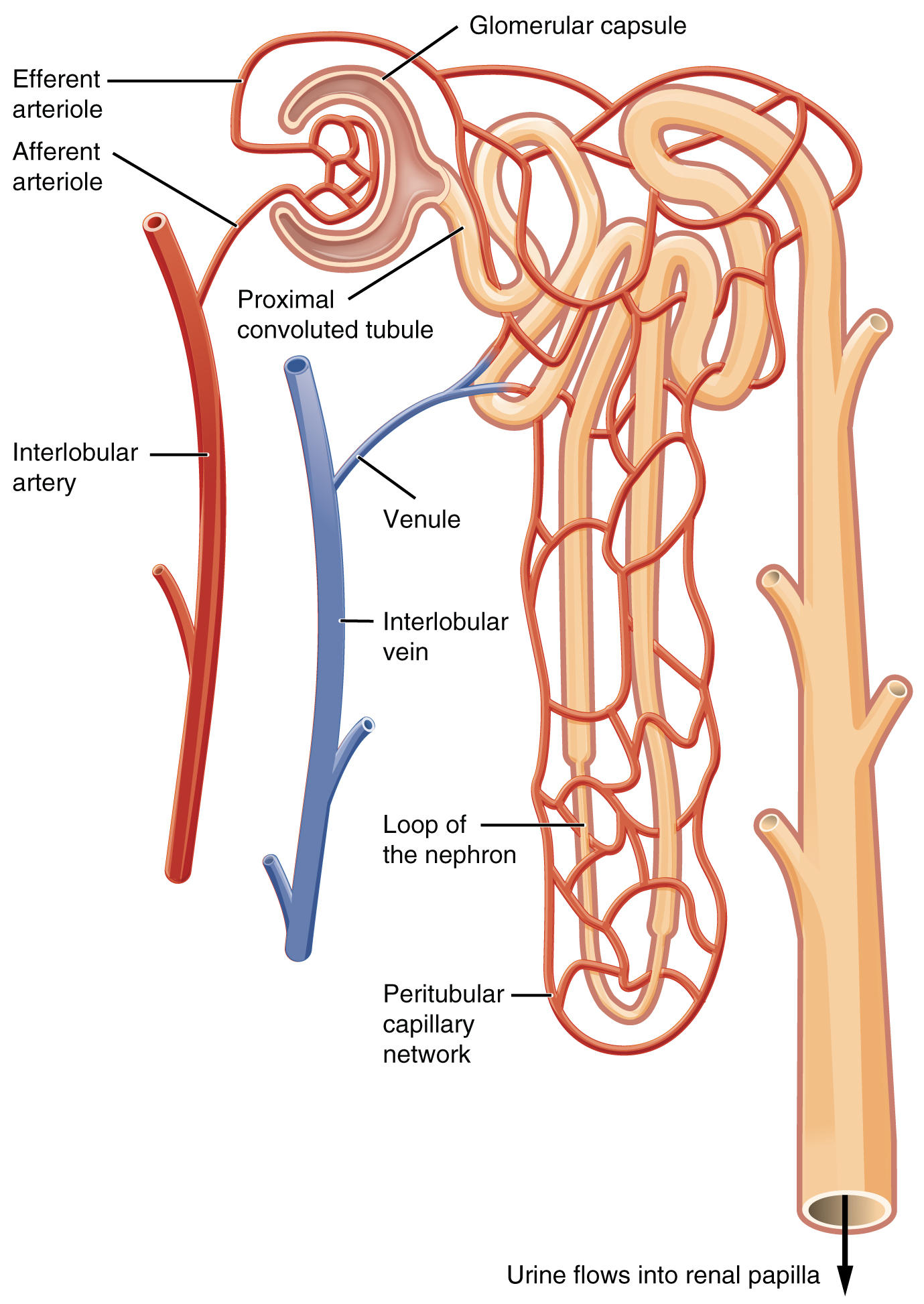 Enlarge illustration ↗
Trace two separate routes: blood passes through the glomerulus and then the capillaries around the tubule; tubular fluid follows the tubule toward the collecting duct. Their close contact allows exchange without mixing the two fluids.
Image source and licence
OpenStax College · Source image · CC BY 3.0. Original image, reproduced without alteration.
Cortical and juxtamedullary nephrons
Not all nephrons are identical. Cortical nephrons, which make up the majority, have their corpuscles in the outer part of the cortex and relatively short loops of Henle that barely enter the medulla. Juxtamedullary nephrons have their corpuscles close to the medulla and long loops that descend deep into it. This difference matters because the ability to produce concentrated urine depends on the long loops and their associated vasa recta, which build up a gradient of solutes in the medulla. Animals that must conserve water, such as desert species, have a high proportion of juxtamedullary nephrons with very long loops. In humans, the same principle applies: the longer the loops, the more water can be reclaimed and the more concentrated the urine can become.
For example: A desert rodent can produce urine far more concentrated than human urine because its loops of Henle are much longer.
Key facts to remember
- The urinary organs are the two kidneys, two ureters, the urinary bladder and the urethra.
- The kidney has an outer cortex, an inner medulla divided into renal pyramids, and a central renal pelvis that continues as the ureter.
- The nephron has a renal corpuscle (glomerulus plus glomerular capsule) and a renal tubule (proximal tubule, loop of Henle, distal tubule and collecting duct).
- The glomerulus is a high-pressure capillary bed between an afferent and a narrower efferent arteriole, followed by peritubular capillaries and vasa recta.
- Juxtamedullary nephrons have long loops that descend into the medulla and allow concentrated urine to be produced.
Example · Follow the path of a drop of urine from its formation to the outside. Fluid is filtered from the glomerulus into the glomerular capsule in the cortex. It flows through the proximal convoluted tubule, down and up the loop of Henle into the medulla and back, through the distal convoluted tubule, and into a collecting duct. The collecting duct carries it through a renal pyramid to a calyx, then into the renal pelvis. From there it enters the ureter, is stored in the urinary bladder, and leaves through the urethra. Cortex to medulla, pelvis to ureter, bladder to urethra.
Read full notes
Read one topic at a time. These notes explain the mechanisms and relationships behind the key facts.
The urinary organs and their positions
The organs of the urinary system are arranged so that urine moves by gravity and by muscular contraction from where it is made to where it is stored and expelled. The kidneys are retroperitoneal, lying behind the peritoneal cavity against the posterior abdominal wall, which is why they are protected by the lower ribs and why their pain is felt in the flank. The ureters run downward and inward to the bladder, and their walls contain smooth muscle that produces waves of contraction called peristalsis, so urine moves even when a person is lying down. The bladder is a reservoir with a muscular wall, and the urethra is the exit tube. This simple arrangement allows urine production to continue continuously while emptying occurs intermittently.
Inside the kidney: cortex, medulla and pelvis
The internal organisation of the kidney is a map of its functions. The cortex houses the filters, the medulla houses the machinery that concentrates urine, and the pelvis collects the finished product. The pyramids are the visible expression of the medullary machinery: their broad bases face the cortex and their tips, called papillae, point into the calyces. Each papilla delivers urine into a calyx, and the calyces join to form the pelvis. Because concentrating urine requires a solute gradient in the medulla, the lengths of the loops of Henle and the arrangement of the vasa recta are critical. A kidney that loses its medullary organisation, as in some chronic diseases, loses the ability to concentrate urine, and the person produces large volumes of dilute urine.
The nephron: corpuscle and tubule
The nephron is often described in two halves because its two halves do different jobs. The renal corpuscle is the filtration unit: its glomerulus is a high-pressure capillary bed, and its capsule collects the fluid that is forced out. Nothing is selectively removed at this stage; filtration is driven by pressure and depends mainly on the size and charge of molecules. The renal tubule is the modification unit: as filtrate passes along it, useful substances are reabsorbed into the peritubular capillaries and waste products are secreted into the tubule. By the time the filtrate reaches the collecting duct, it has become urine. Understanding this division makes the rest of renal physiology easier, because each question about the kidney can be traced to either filtration in the corpuscle or modification in the tubule.
Blood supply of the nephron
The renal circulation is unusual because it contains two capillary beds in series. Blood passes first through the glomerulus, where filtration occurs, and then through the peritubular capillaries or vasa recta, where exchange with the tubule occurs. Two features make this work. First, the efferent arteriole is narrower than the afferent arteriole, so pressure remains high in the glomerulus; if this balance changes, filtration changes with it. Second, the second capillary bed is arranged around the tubule so that reabsorbed substances can be returned to the blood immediately and secreted substances can be delivered to the tubule. This design links filtration, reabsorption and secretion into one continuous circuit and explains why changes in blood pressure affect urine formation so directly.
Cortical and juxtamedullary nephrons
The two types of nephron are a good example of structure matching environment. Cortical nephrons perform the routine work of filtration and reabsorption and are numerous. Juxtamedullary nephrons, though fewer, are specialised for water conservation because their long loops and vasa recta generate the medullary gradient. The proportion of each type varies between species according to water availability, and within a species the gradient can be adjusted by hormones. This is why the kidney can produce dilute urine after drinking a large volume of water and concentrated urine during dehydration: the same anatomy is used in different ways. Recognising the two types also explains why damage to the medulla has a greater effect on concentrating ability than damage to the cortex.
Explore the diagram
Start with the whole view, then select a numbered structure. Compare the additional illustrations to connect what a structure looks like with what it does.
Kidneys to bladder
Two kidneys drain through two ureters into one bladder. The urethra is the outlet.
More controls
01 / 6 Right kidney
Right kidney
The kidneys filter plasma and adjust water, salts and acid–base balance. The right often lies slightly lower.
Illustration credits
- Jmarchn · Urinary system.svg · CC BY-SA 3.0.

Adapted for this lesson: labels removed, artwork cropped or recoloured, and interactive teaching overlays added. Adapted artwork retains its source licence.
Follow fluid through a nephron
One simplified nephron with its collecting duct; neighbouring nephrons are omitted. Blood and tubular fluid occupy separate spaces. The cortex is above the dashed boundary.
The glomerular capillaries lie inside a capsule. Filtration moves water and small solutes from blood into the capsular space; cells and most proteins remain in blood.
Connect the illustrations to the key ideas
URINARY ORGANS · kidneys → ureters → bladder → urethra
The kidneys are retroperitoneal in the flanks; the bladder lies in the pelvis.
INSIDE THE KIDNEY · cortex, medulla with pyramids, renal pelvis
Corpuscles lie in the cortex; loops and collecting ducts run through the medulla to the pelvis.
THE NEPHRON · corpuscle → proximal tubule → loop → distal tubule → collecting duct
Blood enters by the afferent arteriole and leaves by the narrower efferent arteriole.
Core Concepts Defined
- Kidney
- A bean-shaped organ that filters blood and produces urine.
- Hilum
- The indented side of the kidney where the artery, vein and ureter enter or leave.
- Ureter
- The muscular tube carrying urine from a kidney to the bladder.
- Urinary bladder
- The hollow muscular sac that stores urine in the pelvis.
- Urethra
- The tube through which urine leaves the body.
- Renal cortex
- The outer region of the kidney, containing the renal corpuscles.
- Renal medulla
- The inner region of the kidney, divided into renal pyramids.
- Renal pyramid
- A triangular structure of the medulla whose tip points toward the pelvis.
- Renal column
- An extension of cortex passing between renal pyramids.
- Renal pelvis
- The funnel-shaped cavity that collects urine and continues as the ureter.
- Calyx
- A cup-like extension of the renal pelvis that receives urine from a pyramid.
- Nephron
- The functional unit of the kidney, comprising a renal corpuscle and a renal tubule.
- Renal corpuscle
- The glomerulus together with its surrounding glomerular capsule.
- Glomerulus
- The tuft of high-pressure capillaries where filtration begins.
- Glomerular (Bowman's) capsule
- The cup-shaped chamber that collects the filtrate from the glomerulus.
- Proximal convoluted tubule
- The coiled first part of the renal tubule, in the cortex.
- Loop of Henle
- The hairpin-shaped tubule segment that dips into the medulla.
- Distal convoluted tubule
- The coiled tubule segment after the loop of Henle, in the cortex.
- Collecting duct
- The duct receiving several nephrons and carrying urine through the medulla to a calyx.
- Afferent arteriole
- The vessel bringing blood into the glomerulus.
- Efferent arteriole
- The narrower vessel carrying blood out of the glomerulus, helping maintain filtration pressure.
- Peritubular capillaries
- The capillary network around the tubule where reabsorbed and secreted substances exchange.
- Vasa recta
- Long vessels running alongside the loop of Henle into the medulla.
- Cortical nephron
- A nephron with its corpuscle in the outer cortex and a short loop of Henle.
- Juxtamedullary nephron
- A nephron with its corpuscle near the medulla and a long loop of Henle.
Go deeper · worked example
Locating each part of the nephron
- Start at the filter. The renal corpuscle lies in the cortex. Blood enters the glomerulus through the afferent arteriole, fluid is filtered into the glomerular capsule, and blood leaves through the efferent arteriole.
- Follow the first coiled segment. Filtrate enters the proximal convoluted tubule, which remains in the cortex and is the site of most reabsorption.
- Dip into the medulla. The loop of Henle descends into the medulla and returns to the cortex; the length of this loop determines how concentrated urine can become.
- Pass the second coiled segment. Filtrate enters the distal convoluted tubule in the cortex, where fine adjustment of solute and pH balance occurs under hormonal control.
- Finish in the collecting duct. Several nephrons drain into one collecting duct, which passes through the medulla to a calyx, then to the renal pelvis, the ureter and the bladder.
Explain it yourself · Explain why the loop of Henle is the only part of the nephron that leaves the cortex.
Reveal a model explanation
The cortex contains the renal corpuscles and the convoluted tubules, and it is where filtration and most reabsorption occur. The loop of Henle descends into the medulla and returns, and its purpose is to create and use the high solute concentration of the medullary tissue. Because the medulla is saltier than the cortex, water can be drawn out of the descending limb and, later, out of the collecting duct as it passes through the same region. This is what allows the kidney to produce urine more concentrated than blood. The longer the loop and the deeper it descends, the greater the concentration gradient it can support, which is why juxtamedullary nephrons with long loops are so important for water conservation.
Check
Answer three questions from this section — one easy, one medium and one hard. If you miss one, you get a fresh set with explanations for the last attempt.
Practise this sectionOpen all questions and filter by difficulty or Bloom levelReferences and review status
OpenStax, Anatomy and Physiology 2e, 25.1 Physical Characteristics of Urine and 25.2 Gross Anatomy of Urine Transport and 25.3 Gross Anatomy of the Kidney; Tortora & Derrickson, Principles of Anatomy and Physiology, 12e, ch. 26. Author review only; medical and French-language expert review pending.
Original teaching schematics. English and French wording is author-reviewed for study; anatomy and French-language expert review is pending. Checks are practice only, not a graded examination.
Practice progress is saved on this device for the current learner.