SECTION 7.6
The thymus and T-cell maturation
Identify the thymus, explain its role in maturing T lymphocytes, and describe how its activity changes with age.
Learn
The thymus is a small organ in the upper chest, behind the sternum and above the heart, and it has one main job: to make T lymphocytes safe and functional. Immature lymphocytes are produced in red bone marrow and travel in the blood to the thymus, where they multiply, learn to recognise foreign antigens presented on the body’s own cells, and are tested so that those reacting against the body are removed. Only a small fraction survives this education and leaves to populate lymph nodes, the spleen and other lymphatic tissues. The thymus is called a primary lymphatic organ because it is where lymphocytes mature, in contrast to secondary organs, where mature lymphocytes do their work. Its activity is greatest in childhood, and it gradually shrinks and is replaced by fat with age.
Key Concepts & Topics
A primary lymphatic organ
Lymphatic organs are divided into two groups by what they do. Primary lymphatic organs are where lymphocytes are produced and become mature: they are the red bone marrow, where all lymphocytes originate, and the thymus, where T lymphocytes mature. Secondary lymphatic organs are where mature lymphocytes meet antigens and carry out immune responses: they include lymph nodes, the spleen, tonsils and other mucosa-associated lymphoid tissue. This division matters because it explains why the thymus looks different from a node and why its removal has different consequences. A lymph node filters lymph and responds to antigens throughout life; the thymus produces a supply of educated T cells, mostly early in life, and does not filter lymph at all.
For example: Red bone marrow and the thymus are primary organs; lymph nodes and the spleen are secondary organs where mature cells respond to antigens.
Location and structure
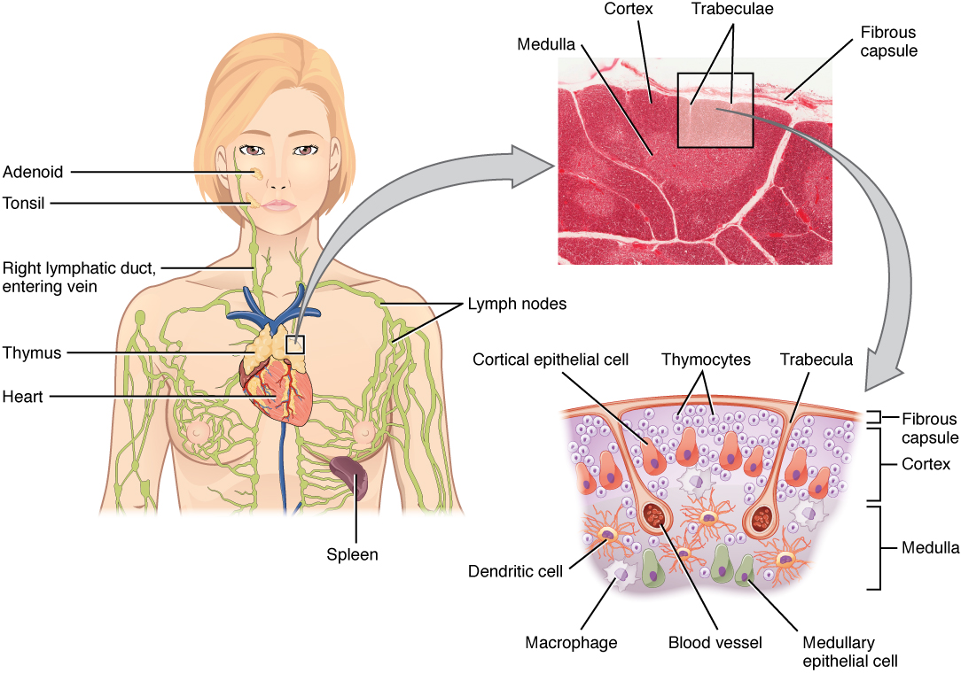
The thymus lies in the superior mediastinum, behind the sternum and in front of the great vessels, extending upward into the lower neck. It has two lobes, each divided into lobules by connective tissue septa. Each lobule has an outer cortex and an inner medulla. The cortex is densely packed with immature T cells, called thymocytes, along with supporting epithelial cells that provide the signals they need to develop. The medulla is paler and contains fewer, more mature thymocytes plus distinctive whorls of epithelial cells called thymic corpuscles. Unlike lymph nodes, the thymus has no afferent lymphatic vessels: cells arrive by blood, not by lymph. It does have blood vessels and a capsule, and its epithelial framework is essential, because developing T cells depend on contact with epithelial cells to survive and mature.
For example: Thymic corpuscles in the medulla are a histological landmark that helps identify thymus tissue under the microscope.
 Enlarge illustration ↗
Move from the chest view to a thymic lobule. The darker cortex and lighter medulla are different regions within the same organ. T cells develop and undergo selection here; the thymus is not a lymph-filtering station.
Image source and licence
OpenStax College · Source image · CC BY 3.0. Original image, reproduced without alteration.
How T cells are selected
T-cell maturation is a process of selection rather than simple growth. Immature lymphocytes arrive from the bone marrow, multiply in the cortex and begin to express receptors that recognise antigens only when they are presented by the body’s own marker molecules, called MHC proteins. They are then tested twice. In positive selection, cells that can recognise the body’s MHC molecules receive a survival signal, while those that cannot are allowed to die. In negative selection, cells whose receptors react strongly against the body’s own tissues are removed, which reduces the risk of autoimmunity. Only a small proportion, often estimated at a few per cent, survives both tests and leaves the thymus as mature, self-tolerant T cells. This education is why the thymus is described as a school for T cells.
For example: If negative selection fails, self-reactive T cells escape into the tissues and can contribute to autoimmune disease.
The thymus through life
The thymus is proportionally largest in infancy and childhood, when the immune system is being established, and it remains active through puberty. After puberty it gradually involutes: the lymphoid tissue is progressively replaced by fatty tissue, and the output of new T cells falls. In an older adult the thymus may be small and largely fatty, yet the person is not left without T-cell immunity, because mature T cells produced earlier continue to circulate and divide, and memory T cells persist for decades. Involution is therefore a normal ageing change, not a disease. It does mean, however, that recovery of T-cell numbers after severe depletion is slower in older people, because the organ that produces new T cells is less active.
For example: A chest X-ray in a child may show a thymic shadow, while in an adult the thymus is usually too small to be seen.
Clinical relevance
Because the thymus is essential for producing T cells, its absence or malfunction has serious consequences, especially early in life. In DiGeorge syndrome, the thymus fails to develop properly, so T-cell numbers are low and the person is vulnerable to infections that would normally be controlled by T cells. The thymus can also become the site of a tumour, called a thymoma, and because thymic tissue is involved in teaching the immune system to tolerate the body, disorders of the thymus are associated with autoimmune conditions such as myasthenia gravis. Surgical removal of the thymus in an adult usually has less dramatic effects than in a newborn, because the adult already has a large pool of mature circulating T cells produced earlier in life.
For example: An infant with DiGeorge syndrome may need specialised care and protective measures because T-cell-mediated defence is impaired.
Key facts to remember
- The thymus is a primary lymphatic organ in the superior mediastinum where T lymphocytes mature.
- Primary lymphatic organs are red bone marrow and the thymus; secondary organs include lymph nodes, the spleen and lymphoid nodules.
- Each thymic lobule has a cortex of immature thymocytes and a medulla with more mature cells and thymic corpuscles.
- Positive selection keeps cells that recognise the body’s MHC; negative selection removes cells that react strongly against self, producing self-tolerant T cells.
- The thymus is largest in childhood and involutes after puberty, replaced largely by fat, though circulating and memory T cells persist.
Example · Trace a T cell from its birth to its work. It originates in red bone marrow and travels in the blood to the thymus, where it enters the cortex as an immature thymocyte. It multiplies and develops a receptor. In positive selection it must recognise the body’s MHC molecules to survive; in negative selection it must not react strongly against the body’s own tissues. If it passes both tests, it leaves the medulla as a mature, self-tolerant T cell and settles in a lymph node, the spleen or mucosa-associated lymphoid tissue, ready to recognise antigens presented by other cells. Most cells do not pass, which is why the thymus is a school with a low graduation rate.
Read full notes
Read one topic at a time. These notes explain the mechanisms and relationships behind the key facts.
A primary lymphatic organ
The distinction between primary and secondary lymphatic organs clarifies how the immune system is organised. Primary organs, red bone marrow and thymus, are the sites of lymphocyte production and maturation; they do not respond to antigens. Secondary organs, lymph nodes, spleen, tonsils and other mucosa-associated lymphoid tissue, are where mature lymphocytes meet antigens and where immune responses are generated. This means the thymus is not a filter and not a site of antibody production; it is a school. Its product, a diverse population of self-tolerant T cells, is exported to the secondary organs, where it does its work. Understanding the division also explains why the thymus is active early in life and why its role changes with age.
Location and structure
The thymus sits where it can be supplied with immature cells from the blood and where it is protected: in the superior mediastinum behind the sternum. Its structure supports its educational role. The cortex, packed with immature thymocytes, is where cells multiply and begin to develop receptors. The medulla, with more mature cells and thymic corpuscles, is where the final stages of selection and export occur. Epithelial cells run through both regions and are not merely structural: they provide the signals, including MHC molecules, that immature T cells need in order to be tested. The absence of afferent lymphatic vessels reinforces the point that the thymus receives cells, not lymph; it is supplied by blood vessels and drained by lymph leaving it.
How T cells are selected
Selection solves two problems at once. The immune system must produce T cells able to recognise antigens presented by the body's own MHC molecules, otherwise they would be useless; and it must avoid producing T cells that attack the body, otherwise they would cause autoimmune disease. Positive selection keeps cells that can interact with MHC, ensuring the receptor is useful. Negative selection removes cells that bind self-antigens too strongly, ensuring the receptor is safe. Because both tests are stringent, most developing cells die in the thymus, and only a small proportion is exported. This apparent waste is the price of a system that must generate enormous diversity while remaining tolerant of the body itself.
The thymus through life
The life history of the thymus explains several clinical observations. Because it is most active early, conditions that damage it in infancy have severe effects on T-cell immunity; because it involutes later, the same damage in adulthood is better tolerated. Because it shrinks with age, older people regenerate T cells more slowly after illness or treatment that depletes them. Yet involution does not mean the loss of T-cell immunity: mature T cells already in the circulation continue to divide, and memory T cells persist for decades. The thymus is therefore best understood as the organ that builds the T-cell repertoire early and then gradually hands over the job of maintaining it to the cells it has already produced.
Clinical relevance
Thymic disorders illustrate the organ’s importance. When the thymus fails to develop, as in DiGeorge syndrome, T-cell numbers are low and infections that require T-cell help are poorly controlled. When the thymus develops a tumour, a thymoma, it may cause local effects and is also associated with autoimmune conditions, reflecting the organ’s role in teaching self-tolerance. Because the thymus is active early and involutes later, the timing of any insult matters greatly: the same event that is catastrophic in a newborn may be relatively well tolerated by an adult who already possesses a broad repertoire of mature and memory T cells. This is why neonatal thymic function is treated as a priority in clinical assessment.
Explore the diagram
Start with the whole view, then select a numbered structure. Compare the additional illustrations to connect what a structure looks like with what it does.
Thymic lobules: cortex around medulla
Enlarged lobules, not a whole-body view. The thymus lies behind the sternum and is most active early in life; its tissue is increasingly replaced by fat with age.
The darker outer region contains many immature T cells. Their development includes selection for useful recognition of the body’s MHC molecules.
Connect the illustrations to the key ideas
LOCATION AND STRUCTURE · superior mediastinum; lobules with cortex and medulla
It has no afferent lymphatics; immature cells arrive by blood.
SELECTION · positive selection for MHC, negative selection against self
Most thymocytes die; survivors leave as mature self-tolerant T cells.
CHANGES WITH AGE · largest in childhood, involutes after puberty
Fat replaces lymphoid tissue, but circulating and memory T cells persist.
Core Concepts Defined
- Thymus
- A primary lymphatic organ in the superior mediastinum where T lymphocytes mature.
- Primary lymphatic organ
- An organ where lymphocytes are produced or mature: red bone marrow and thymus.
- Secondary lymphatic organ
- An organ where mature lymphocytes encounter antigens: nodes, spleen, nodules.
- Thymic lobule
- A subdivision of a thymic lobe, with an outer cortex and inner medulla.
- Thymic cortex
- The outer region of a lobule, densely packed with immature thymocytes.
- Thymic medulla
- The inner region of a lobule, with more mature thymocytes and thymic corpuscles.
- Thymic corpuscle (Hassall's corpuscle)
- A whorl of epithelial cells in the thymic medulla, a histological landmark.
- Thymocyte
- An immature T lymphocyte developing in the thymus.
- Positive selection
- Selection that keeps thymocytes able to recognise the body’s MHC molecules.
- Negative selection
- Selection that removes thymocytes reacting strongly against the body’s own tissues.
- MHC protein
- A body marker molecule that presents antigens to T cells.
- Self-tolerance
- The state in which T cells do not react against the body’s own tissues.
- Thymic involution
- The age-related shrinking of the thymus and its replacement by fat.
- Thymoma
- A tumour arising from the thymus.
- DiGeorge syndrome
- A condition in which the thymus develops abnormally, impairing T-cell immunity.
Go deeper · worked example
Why the thymus matters more in a newborn
- Identify what the thymus produces. It is the organ where immature lymphocytes from bone marrow become mature, self-tolerant T cells.
- Ask what the newborn has. A newborn has had little time for mature T cells to accumulate, so the supply of functional T cells depends heavily on the thymus being active.
- Ask what the adult has. An adult has decades of circulating mature T cells and long-lived memory T cells produced earlier, so the loss of new production is partly buffered.
- Consider the involution. After puberty the thymus shrinks and its output falls, so even in health the reliance on new thymic production declines with age.
- Conclude. Removing or losing thymic function in a newborn severely limits the development of T-cell immunity, while in an adult the existing pool of mature and memory T cells softens the effect, although the ability to generate new T cells is reduced.
Explain it yourself · Explain why the thymus is called a primary lymphatic organ and why its size decreases with age.
Reveal a model explanation
It is called a primary lymphatic organ because it is one of the two sites where lymphocytes mature, together with red bone marrow. Immature T cells arrive from the marrow, are tested by positive and negative selection, and leave as mature, self-tolerant cells. Lymph nodes, the spleen and lymphoid nodules are secondary organs because that is where mature lymphocytes meet antigens and respond. The thymus is large in childhood because the immune system is being built and large numbers of new T cells are needed. After puberty, the supply of new T cells becomes less critical because a long-lived pool of mature and memory T cells already exists, so the thymus involutes and is gradually replaced by fat. This shrinking is a normal ageing change, not a disease.
Check
Answer three questions from this section — one easy, one medium and one hard. If you miss one, you get a fresh set with explanations for the last attempt.
Practise this sectionOpen all questions and filter by difficulty or Bloom levelReferences and review status
OpenStax, Anatomy and Physiology 2e, 21.1 Anatomy of the Lymphatic and Immune Systems; Tortora & Derrickson, Principles of Anatomy and Physiology, 12e, T12-22A. Author review only; medical and French-language expert review pending.
Original teaching schematics. English and French wording is author-reviewed for study; anatomy and French-language expert review is pending. Checks are practice only, not a graded examination.
Practice progress is saved on this device for the current learner.