SECTION 7.5
The spleen: blood surveillance and storage
Relate the structure of the spleen’s red and white pulp to its roles in blood surveillance, blood-cell removal and platelet storage.
Learn
The spleen is the largest lymphatic organ, about the size of a clenched fist, tucked under the ribs on the left side of the abdomen behind the stomach. Unlike a lymph node, it is not placed on a lymphatic vessel; it is inserted into the bloodstream, and it filters blood rather than lymph. Inside, the spleen is divided into two functionally different regions. White pulp is lymphatic tissue built around small arteries, where lymphocytes monitor the blood for antigens. Red pulp is a spongy network of blood-filled channels and macrophages, where worn-out red blood cells and microbes are removed and where platelets are stored. This section explains the structure of the spleen, how the two pulps carry out their different jobs, and what happens when the spleen is removed or overactive.
Key Concepts & Topics
A blood filter, not a lymph filter
The spleen differs from lymph nodes in what it filters. Lymph nodes receive lymph through afferent vessels; the spleen receives blood through the splenic artery and returns it through the splenic vein. This means the spleen inspects the blood itself, including red blood cells, platelets and any microbes that have entered the circulation. Its position in the bloodstream explains its main jobs: it removes aged or damaged red blood cells, it recycles the iron and other components of haemoglobin, it stores a proportion of the body’s platelets, and it mounts immune responses to antigens found in blood. Because blood passes through the spleen under pressure, the organ is well supplied with vessels and is soft and highly vascular, which also makes it liable to bleed if injured.
For example: In a severe blow to the left upper abdomen, the spleen can rupture and bleed heavily, which is why it is protected by the lower ribs.
White pulp: immune surveillance of blood
White pulp is the lymphatic part of the spleen. It is organised as cylindrical sheaths of lymphocytes around the small arteries that branch through the organ, with nodules of B cells at intervals. Because the sheaths surround arteries, lymphocytes are positioned to meet antigens the moment they arrive in the blood. When a blood-borne microbe appears, dendritic cells and macrophages capture it, T cells in the sheath respond, and B cells in the nodules multiply and produce antibodies. This is why the spleen is particularly important in defence against encapsulated bacteria that spread in blood, and why people without a functioning spleen are more vulnerable to these infections. White pulp is named for its paler appearance in fresh tissue, in contrast to the blood-filled red pulp around it.
For example: A patient who has had a splenectomy is advised to be vaccinated against encapsulated bacteria such as pneumococcus.
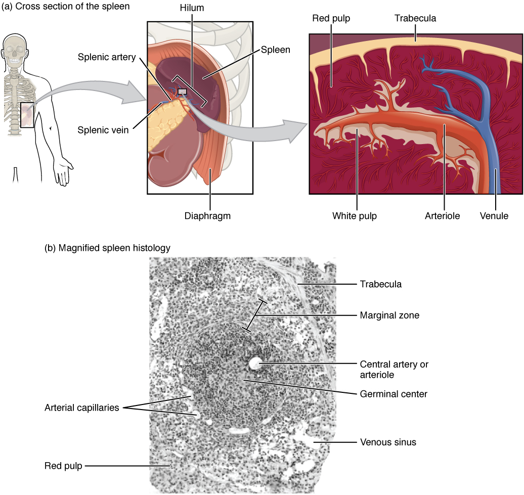 Enlarge illustration ↗
Compare the cutaway with the tissue image. White pulp contains lymphoid tissue involved in immune surveillance; red pulp helps filter blood and remove ageing red cells. Unlike a lymph node, the spleen monitors blood rather than filtering incoming lymph.
Image source and licence
OpenStax College · Source image · CC BY 3.0. Original image, reproduced without alteration.
Red pulp: removing old blood cells
Red pulp fills most of the spleen and is where blood is filtered. It consists of splenic cords, loose networks of reticular fibres, macrophages and blood cells, and of splenic sinuses, the channels through which blood re-enters the venous circulation. Blood flows from the arteries into the cords, where it moves slowly among macrophages. These macrophages test red blood cells as they pass: cells that are old, damaged or abnormal are engulfed and broken down. The iron from their haemoglobin is recovered and returned to the blood for reuse in new red cells, and the remaining components are processed. Because blood cells must squeeze through narrow slits to re-enter the sinuses, rigid or misshapen cells are held back and removed, which is why the spleen is especially effective at clearing abnormal red cells.
For example: In hereditary spherocytosis, red cells are abnormally rigid and spherical, so the spleen removes them early and the person becomes anaemic.
Platelet storage and blood volume
The spleen holds a reserve of platelets and, to a lesser extent, of red blood cells. Roughly a third of the body’s platelets are sequestered in the spleen at any time, held in the red pulp and released into the circulation when needed. The organ also contains a small reservoir of blood that can be mobilised during bleeding or exercise. This storage function has clinical consequences in both directions. If the spleen enlarges, it holds more platelets and blood cells, so their numbers in circulating blood fall. If the spleen is removed, platelet and sometimes red-cell counts rise, because the sequestering and removal functions are gone. Because an enlarged spleen can hold a large proportion of the blood volume, it can also cause a fall in circulating volume and, in extreme cases, circulatory problems.
For example: A patient with an enlarged spleen may have a low platelet count on a blood test even though the bone marrow is producing platelets normally.
When the spleen is injured, removed or overactive
Because the spleen is soft, vascular and close to the ribs, it can be injured in abdominal trauma and bleed extensively; an unstable patient may need surgery to control the bleeding and may lose the organ. Removing the spleen, called splenectomy, is also performed in some blood disorders in which the spleen destroys too many blood cells. After splenectomy, the person has higher platelet counts and a greater risk of infection by encapsulated bacteria, so vaccination and sometimes preventive antibiotics are used. In some conditions the spleen becomes overactive, a state called hypersplenism, and removes too many blood cells, causing anaemia, low platelet counts and increased susceptibility to infection. In each case, the clinical picture follows directly from the spleen’s normal functions.
For example: After a splenectomy, a patient is advised to report fever promptly, because infections can progress rapidly without the spleen’s blood-filtering defence.
Key facts to remember
- The spleen is the largest lymphatic organ and filters blood, not lymph, receiving blood through the splenic artery.
- White pulp is lymphatic tissue arranged around small arteries, where lymphocytes respond to blood-borne antigens.
- Red pulp contains cords and sinuses with macrophages that remove aged or abnormal red blood cells and recycle iron.
- The spleen stores about a third of the body’s platelets and a small reservoir of blood.
- After splenectomy, platelet counts rise and the risk of infection by encapsulated bacteria increases; an overactive spleen removes too many blood cells.
Example · Follow a worn-out red blood cell. It travels in the splenic artery and reaches the red pulp, where blood slows among the splenic cords. A macrophage recognises it as old or damaged and engulfs it. The iron from its haemoglobin is recovered and returned to the blood for reuse, while the rest is broken down. Meanwhile, in the white pulp, lymphocytes around the arteries are sampling the same blood for antigens. If a microbe appears, they mount an antibody response. One organ, two pulps, two filters working on the same blood.
Read full notes
Read one topic at a time. These notes explain the mechanisms and relationships behind the key facts.
A blood filter, not a lymph filter
The spleen is often grouped with lymph nodes because both contain lymphocytes and both filter fluid, but the fluid is different. Nodes filter lymph and therefore inspect what has drained from the tissues; the spleen filters blood and inspects what is circulating in the vessels. This difference shapes everything about the organ. Because blood arrives under arterial pressure and contains red cells and platelets as well as microbes, the spleen needs a robust vascular architecture and a large population of macrophages. It also means the spleen is at risk from the very blood it filters: in conditions where red cells are abnormal, the organ works harder and may enlarge, and in trauma it can bleed heavily. Its functions all follow from its position in the bloodstream.
White pulp: immune surveillance of blood
White pulp is arranged around arteries for a reason: antigens that arrive in the blood are delivered directly to the lymphocytes that must respond to them. The sheaths of T cells and the nodules of B cells form a complete immune apparatus inside the bloodstream. This is why the spleen is essential against organisms that spread in blood, particularly those with capsules that resist simple phagocytosis and require antibodies for efficient clearance. The structure also explains why the immune and filtering functions are separated: white pulp handles recognition and response, while red pulp handles removal and recycling. Keeping them distinct allows each to be optimised, and it means that a problem in one function can occur without the other.
Red pulp: removing old blood cells
The red pulp is a remarkable quality-control system. Blood does not flow through it in sealed pipes; it spills into the cords, where it slows and comes into close contact with macrophages. As red cells re-enter the sinuses, they must pass through narrow slits, so cells with reduced flexibility, such as aged cells or cells with abnormal membranes, are held back and removed. This mechanical testing is why the spleen is so effective at clearing abnormal red cells, and why diseases that make red cells rigid cause them to be destroyed early. The recovered iron is returned to the blood and used again, which makes the spleen an important part of iron recycling as well as of blood-cell quality control.
Platelet storage and blood volume
Storage is an easily overlooked function with clear clinical effects. Because the spleen holds a substantial fraction of the platelets, its size influences the platelet count measured in blood. An enlarged spleen holds more and the count falls; a removed spleen holds none and the count rises. The same logic applies to red cells, though to a smaller degree. Clinicians therefore interpret blood counts with the spleen in mind, and an unexpected combination of low platelets and low red cells may point to the spleen rather than to a problem with production in the marrow. The spleen also holds a small volume of blood that can be released during bleeding or exercise, contributing to the body’s reserve.
When the spleen is injured, removed or overactive
The clinical problems of the spleen are direct translations of its functions. Because it is vascular and close to the ribs, trauma can cause serious bleeding, and an unstable patient may need surgery. Because it removes and stores blood cells, its overactivity lowers their numbers, and its removal raises them. Because its white pulp defends against blood-borne encapsulated bacteria, its loss increases the risk of serious infection. These consequences are not separate diseases but different faces of the same organ. Understanding the two pulps therefore allows a clinician to predict what will happen when the spleen is injured, removed, enlarged or overactive, and to explain the reasoning to a patient.
Explore the diagram
Start with the whole view, then select a numbered structure. Compare the additional illustrations to connect what a structure looks like with what it does.
Two kinds of pulp in the spleen
Schematic cutaway and enlarged tissue regions. The spleen monitors blood, unlike a lymph node which receives and filters lymph.
A connective-tissue covering and inward trabeculae support the spleen. It lies in the upper left abdomen, near the stomach.
Connect the illustrations to the key ideas
SPLEEN IN THE BLOODSTREAM · splenic artery in, splenic vein out
It filters blood rather than lymph, unlike a lymph node.
WHITE PULP · lymphocytes arranged around small arteries
It samples blood-borne antigens and mounts antibody responses.
RED PULP · cords and sinuses with macrophages
It removes aged or abnormal red cells, recycles iron and stores platelets.
Core Concepts Defined
- Spleen
- The largest lymphatic organ, which filters blood and mounts immune responses to blood-borne antigens.
- Splenic artery
- The artery that supplies blood to the spleen.
- Splenic vein
- The vein that carries blood away from the spleen.
- White pulp
- The lymphatic part of the spleen, arranged around small arteries, where lymphocytes respond to blood antigens.
- Red pulp
- The blood-filtering part of the spleen, containing cords and sinuses with macrophages.
- Splenic cord
- A loose network of reticular fibres, macrophages and blood cells in the red pulp.
- Splenic sinus
- A channel in the red pulp through which blood re-enters the venous circulation.
- Haemoglobin recycling
- Recovery of iron and other components from broken-down red blood cells in the spleen.
- Platelet sequestration
- Storage of a proportion of circulating platelets in the spleen.
- Splenectomy
- Surgical removal of the spleen.
- Splenomegaly
- Enlargement of the spleen.
- Hypersplenism
- An overactive spleen that removes excessive numbers of blood cells.
- Encapsulated bacteria
- Bacteria with a polysaccharide capsule, against which the spleen is especially important.
Go deeper · worked example
Why removing the spleen changes the blood count
- Recall the spleen’s blood route. Blood enters through the splenic artery, passes through the red pulp and returns through the splenic vein, so all circulating cells pass through the organ.
- Identify the removal function. In the red pulp, macrophages engulf aged, damaged or abnormally shaped red blood cells, so the spleen normally reduces the number of circulating red cells.
- Identify the storage function. The red pulp also holds about a third of the body’s platelets, so the spleen normally keeps a proportion of platelets out of circulation.
- Predict the effect of removal. Without the spleen, neither removal nor sequestration occurs, so platelet counts rise and red-cell counts may also rise; abnormal red cells may persist in the blood.
- Add the immune consequence. Without white pulp, blood-borne encapsulated bacteria are less efficiently cleared, so the risk of serious infection rises and vaccination plus prompt treatment of fever become important.
Explain it yourself · Explain why a person who has had a splenectomy has a high platelet count and a greater risk of certain infections.
Reveal a model explanation
The spleen normally performs two blood-related jobs. In the red pulp, macrophages remove aged or abnormal red blood cells, and the same red pulp sequesters about a third of the body’s platelets. Removing the spleen therefore removes both the destruction and the storage of these cells, so platelet counts rise and red-cell counts may rise too. In the white pulp, lymphocytes arranged around small arteries monitor the blood for antigens and mount antibody responses, which is especially important against encapsulated bacteria such as pneumococcus. Without the spleen, these organisms are cleared less efficiently and can cause overwhelming infection. This is why patients are vaccinated and advised to seek help quickly for fever.
Check
Answer three questions from this section — one easy, one medium and one hard. If you miss one, you get a fresh set with explanations for the last attempt.
Practise this sectionOpen all questions and filter by difficulty or Bloom levelReferences and review status
OpenStax, Anatomy and Physiology 2e, 21.2 Barrier Defenses and the Innate Immune Response and 21.1 Anatomy of the Lymphatic and Immune Systems; Tortora & Derrickson, Principles of Anatomy and Physiology, 12e, T12-22A. Author review only; medical and French-language expert review pending.
Original teaching schematics. English and French wording is author-reviewed for study; anatomy and French-language expert review is pending. Checks are practice only, not a graded examination.
Practice progress is saved on this device for the current learner.