SECTION 1.5
Surface regions of the body
Locate the main regional and surface landmarks used to describe where a structure or symptom is found.
Learn
Long before anyone studies deep anatomy, the body can be divided by what you can see and feel. Regional terms name areas of the body — the head, neck, trunk and limbs and their subdivisions — while surface landmarks are features you can see, press or measure, such as the breastbone, the collar bone, the ridge of the hip or the kneecap. Together they give a rapid, shared way to say where something is. A rash “over the left shoulder blade”, a pulse “at the front of the wrist” and a pain “below the right rib margin” are all descriptions built from regions and landmarks. This section gives you that vocabulary and, more importantly, teaches you to confirm each landmark on a real body rather than only on a diagram.
Key Concepts & Topics
Axial and appendicular regions
The body is first divided into two great regions. The axial region is the central axis: the head, the neck and the trunk. The trunk contains the thorax (chest), the abdomen and the pelvis. The appendicular region is made of the appendages, the upper limbs and the lower limbs, including the girdles that attach them to the trunk. This split is not just tidiness: the axial region protects the brain, heart, lungs and abdominal organs, while the appendicular region provides movement and manipulation. Many clinical descriptions begin by naming which of the two regions is involved, because the causes and consequences differ. A pain in the axial region may concern a deep organ; a pain in the appendicular region often concerns a joint, muscle or bone.
For example: A fractured wrist is an appendicular problem; chest pain from a heart problem is axial. The region guides the first questions.
Named regions of the trunk and limbs
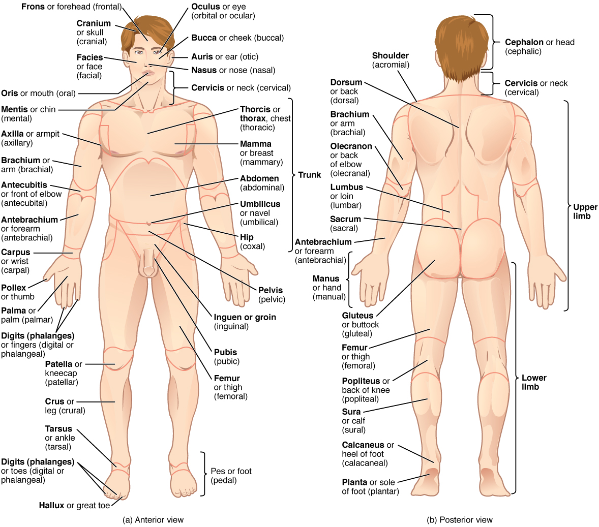
Anatomical regions have standard names. The head is the cephalic region, the neck the cervical region, the chest the thoracic region. The abdomen is the abdominal region and the lower trunk the pelvic region. The back is divided into the vertebral region along the spine and the dorsal region over the shoulder blades. On the upper limb, the arm between shoulder and elbow is the brachial region, the forearm the antebrachial region, and the wrist the carpal region. On the lower limb, the thigh is the femoral region, the leg between knee and ankle the crural region, and the ankle and foot the tarsal and pedal regions. The armpit is the axillary region and the groin the inguinal region. These names let a clinician say exactly which part is meant without listing muscles or bones.
For example: “Brachial” means the arm between shoulder and elbow, not the whole upper limb. Saying “the brachial region” avoids the everyday confusion between arm and forearm.
Common misconception: In anatomy, “arm” is only the segment from shoulder to elbow; the whole limb is the upper limb, and the forearm is a separate region.
 Enlarge illustration ↗
Compare the front and back of the same body. Find the shoulder, elbow and knee in each view; a regional name identifies an area, not one isolated organ.
Image source and licence
Connexions / OpenStax · Source image · CC BY 4.0. Original image, reproduced without alteration.
Surface landmarks you can see and feel
A landmark is a structure you can identify on the surface, and it acts as an anchor for description and for procedures. Common examples include the clavicle (collar bone) across the top of the chest, the sternum (breastbone) in the midline, the ribs and the costal margin where they end, the umbilicus (navel) in the centre of the abdomen, the iliac crest felt at the top of the hip, the anterior superior iliac spine at its front corner, the patella (kneecap), and the spinous processes felt as a ridge down the back. On the posterior knee the hollow is the popliteal region, and at the front of the elbow the cubital fossa is where vessels and nerves lie close to the surface. Landmarks are reliable because they do not change with body weight as much as the soft tissue around them.
For example: The anterior superior iliac spine is a reliable landmark for finding the level of a lumbar puncture or for locating the appendix.
Using landmarks to describe a finding
A useful description names the side, the region and at least one landmark, then adds size, colour or depth as needed. “A rash on the right upper back, over the medial border of the shoulder blade” is far more useful than “a rash on the back”. “A pulse felt at the anterior wrist, lateral to the tendon in the middle” locates the radial pulse precisely. Landmarks also let a clinician measure: the distance from the sternal notch to a wound, or the height of a swelling above the patella, is meaningful because the anchor is fixed. When you describe a symptom, imagine the next person reading your note without seeing the patient; if they could place a finger on the same spot, the description is complete enough.
For example: “Swelling 3 cm above the right patella” is repeatable by another clinician, because the kneecap is a fixed anchor.
Why surface anatomy matters in care
Surface anatomy guides many everyday procedures and observations. Taking a pulse means knowing where an artery lies close to the surface and over a firm base, such as the radial, brachial, carotid, femoral and popliteal pulses. Giving an injection safely means recognising regions with few vital structures at the chosen depth, and avoiding nerves and vessels. Listening to the chest means placing the stethoscope over known lung and heart regions rather than at random. Measuring blood pressure means finding the brachial artery and wrapping the cuff at the right level. In each case the skill depends on being able to find a landmark quickly by touch and to translate what you feel into a precise description.
For example: The brachial pulse is felt on the medial side of the upper arm, between the biceps and the humerus, which is why the blood-pressure cuff is placed there.
Key facts to remember
- The axial region is the head, neck and trunk; the appendicular region is the upper and lower limbs with their girdles.
- Standard regional names include cephalic (head), cervical (neck), thoracic (chest), abdominal, pelvic, brachial (arm), antebrachial (forearm), femoral (thigh) and crural (leg).
- Landmarks such as the sternum, clavicle, costal margin, umbilicus, iliac crest and patella anchor a description to fixed points.
- A precise description names the side, the region and a landmark, then adds size, colour or depth.
- Surface anatomy guides pulse taking, injections, auscultation and blood-pressure measurement.
Example · A patient reports pain at the front of the right hip. The nurse names the side (right), the region (hip, part of the pelvic/appendicular junction) and a landmark (the anterior superior iliac spine). Examining further, the pain is 2 cm medial and inferior to that spine, over the groin. The full description — “right-sided pain just medial and inferior to the anterior superior iliac spine, in the inguinal region” — allows another clinician to examine exactly the same place.
Read full notes
Read one topic at a time. These notes explain the mechanisms and relationships behind the key facts.
Two great regions and what they do
The axial and appendicular division reflects function as well as position. The axial skeleton protects the most vital and least replaceable structures — the brain, spinal cord, heart and lungs — and the trunk contains the organs of digestion, excretion and reproduction. The appendicular region specialises in movement, reach and manipulation, and its joints are built for range rather than protection. Because the two regions have different jobs, injuries affect them differently. A blow to the appendicular region often produces a joint or muscle injury that the patient can still move around; a blow to the axial region can injure organs with no external wound at all. Naming the region first is therefore a way of asking the right next question.
Learning the regional vocabulary
Regional terms often come from Latin or Greek roots, and the roots repeat. Cephalic comes from the Greek for head, cervical from the Latin for neck, brachial from the Latin for arm, femoral from the Latin for thigh. Learning a few roots makes many names predictable: “-al” usually means “relating to”, so thoracic means relating to the thorax, and axillary means relating to the axilla. Regions are also nested: the upper limb contains the brachial, antebrachial and carpal regions, and the trunk contains the thoracic, abdominal and pelvic regions. When you learn a new term, place it in the hierarchy rather than memorising it in isolation. This makes recall easier and prevents the common mistake of using a regional term for a larger area than it actually covers.
Landmarks, and how to confirm them
A landmark is only useful if you can find it reliably. Practise on yourself and on others: run a finger along the clavicle from the sternum outward until you feel its lateral end; press along the front of the hip to find the iliac crest, then roll forward to its front corner, the anterior superior iliac spine; feel the patella and the tibial tuberosity just below it; follow the spinous processes down the back. Landmarks are chosen because they are both visible or palpable and consistent between people of different sizes. When a landmark is hard to find, the reason is usually soft tissue: fat, muscle or oedema can bury a structure that is obvious in a lean person. Recording which landmark you used lets a colleague repeat the examination even if their own palpation differs slightly.
From landmark to measurement
Landmarks turn descriptions into measurements that can be compared over time. Instead of “the swelling is large”, say “the swelling extends 4 cm above the superior border of the patella”. Instead of “the wound is near the elbow”, say “the wound lies 3 cm distal to the olecranon on the posterior forearm”. Measured from a fixed anchor, the value is meaningful in a body of any size and can be compared with the next observation. This is why wound charts, pressure-injury records and many assessment forms ask for distances from a named landmark rather than for an impression. Accurate landmarks make the difference between a note that can be audited and one that must be guessed at.
Surface anatomy in procedures
Many procedures are safe because of surface anatomy. Pulse points are chosen where an artery passes over bone, so the pulsation is not cushioned by muscle. Injections into a muscle are placed in a region with a predictable thickness and few vital structures, and the site is chosen to avoid nerves and large vessels. A cannula is placed where a vein is visible and supported. Chest auscultation uses fixed surface regions so that a finding can be described as “right lower zone” or “apex”, and compared over time. In every case the professional is using the same two tools: a named region and a palpable landmark. Learning them early converts anatomy from a subject to be memorised into a practical map used every day.
Explore the diagram
Start with the whole view, then select a numbered structure. Compare the additional illustrations to connect what a structure looks like with what it does.
Anterior landmarks
The skeleton is visible through the surface to connect external landmarks with deeper support.
More controls
01 / 7 Clavicle
Clavicle
A palpable bone at the base of the front of the neck.
Illustration credits
- Goran tek-en · Male front 3d-shaded human illustration.svg · CC BY-SA 4.0.

Adapted for this lesson: labels removed, artwork cropped or recoloured, and interactive teaching overlays added. Adapted artwork retains its source licence.
Posterior landmarks
The skeleton is visible through the surface to connect external landmarks with deeper support.
More controls
01 / 6 Occipital region
Occipital region
The back of the head overlies the occipital bone.
Illustration credits
- Goran tek-en · Male back 3d-shaded human illustration.svg · CC BY-SA 4.0.

Adapted for this lesson: labels removed, artwork cropped or recoloured, and interactive teaching overlays added. Adapted artwork retains its source licence.
Connect the illustrations to the key ideas
AXIAL REGION · head, neck and trunk
The central axis contains and protects the brain, heart, lungs and abdominal organs.
APPENDICULAR REGION · upper and lower limbs with their girdles
The limbs provide movement, reach and manipulation; their regions include brachial, antebrachial, femoral and crural.
SURFACE LANDMARKS · clavicle, sternum, costal margin, iliac crest, patella
Visible or palpable anchors let a colleague find the exact same place and repeat a measurement.
Core Concepts Defined
- Axial region
- The central part of the body: head, neck and trunk.
- Appendicular region
- The limbs and the girdles that attach them to the trunk.
- Surface anatomy
- The study of structures that can be seen or felt on the body surface and used as landmarks.
- Landmark
- A visible or palpable structure that anchors a description or a procedure.
- Cephalic
- Relating to the head.
- Cervical
- Relating to the neck.
- Thoracic
- Relating to the chest.
- Abdominal
- Relating to the abdomen, the part of the trunk below the chest and above the pelvis.
- Pelvic
- Relating to the pelvis, the lower part of the trunk.
- Brachial
- Relating to the arm, the segment from shoulder to elbow.
- Antebrachial
- Relating to the forearm, between elbow and wrist.
- Carpal
- Relating to the wrist.
- Femoral
- Relating to the thigh.
- Crural
- Relating to the leg, the segment between knee and ankle.
- Pedal
- Relating to the foot.
- Axillary
- Relating to the armpit.
- Inguinal
- Relating to the groin.
- Umbilicus
- The navel; a midline landmark of the abdomen.
- Iliac crest
- The upper ridge of the hip bone, felt at the side of the waist.
- Popliteal
- Relating to the hollow behind the knee.
Go deeper · worked example
Locating a finding from region and landmark
- Start with the great region. Decide whether the finding is axial (head, neck, trunk) or appendicular (limb). Here, assume a lump in the upper chest wall: it is in the axial region.
- Narrow to the named region. The upper chest is the thoracic region, on the anterior side of the trunk.
- Find a landmark. The clavicle crosses the top of the chest and is easily felt; the sternum lies in the midline.
- Relate the finding to the landmark. Say whether it is medial or lateral to the landmark, and superior or inferior to it. For example, “2 cm lateral to the sternum, just inferior to the clavicle”.
- Add the side and any measurement. State left or right, then size or depth. The final description is repeatable by another clinician without a picture.
Explain it yourself · Describe, in your own words, how you would locate the brachial pulse to a colleague who cannot see the patient.
Reveal a model explanation
The brachial pulse is found on the medial side of the arm, in the brachial region between the shoulder and the elbow, just medial to the biceps muscle and against the humerus. To describe it: the upper limb, the arm segment, the medial surface, about midway between shoulder and elbow, where the artery passes close to the bone over a firm base. Because the description uses region plus surface plus landmark, a colleague can press the same place.
Check
Answer three questions from this section — one easy, one medium and one hard. If you miss one, you get a fresh set with explanations for the last attempt.
Practise this sectionOpen all questions and filter by difficulty or Bloom levelReferences and review status
OpenStax, Anatomy and Physiology 2e, 1.6 Anatomical Terminology (regional and surface terms); Tortora & Derrickson, Principles of Anatomy and Physiology, 12e, ch. 1. Author review only; medical and French-language expert review pending.
Optional further reading · English · internet required:
- Anatomy and physiology: an overview
- Structural organisation of the body
- Homeostasis and feedback
- Anatomical terminology
Original teaching schematics. English and French wording is author-reviewed for study; anatomy and French-language expert review is pending. Checks are practice only, not a graded examination.
Practice progress is saved on this device for the current learner.