SECTION 8.3
Regulating blood volume and pressure
Explain how the kidneys use ADH, aldosterone and the renin–angiotensin–aldosterone system to control blood volume and pressure.
Learn
The kidneys control how much water and sodium leave the body, and because water and sodium determine the volume of the blood, the kidneys are the main long-term regulators of blood pressure. Two hormones do most of the work. Antidiuretic hormone (ADH) controls how much water the collecting ducts reabsorb, so it sets how concentrated the urine is. Aldosterone controls how much sodium is reabsorbed in the distal nephron, and water follows the sodium. Aldosterone is released mainly through the renin–angiotensin–aldosterone system, a chain of signals that begins when the kidney detects low blood pressure or low sodium and ends with vasoconstriction and sodium retention. Other signals, including atrial natriuretic peptide from the heart, work in the opposite direction. This section explains the main pathways and how they keep blood volume and pressure within a workable range.
Key Concepts & Topics
Why volume and pressure need regulation
Blood pressure depends on how much blood the vessels contain and how narrow the vessels are. Because water and sodium are the main determinants of blood volume, the kidneys can raise pressure by retaining them and lower pressure by excreting them. This is a slow, powerful mechanism: adjusting the daily output of water and sodium by even a small amount changes blood volume over hours and days, and it is the reason the kidneys dominate long-term blood pressure control. Short-term adjustments are made by the heart and blood vessels, which can change rate and diameter within seconds, but the balance between intake and output is settled by the kidneys. When the kidneys retain too much sodium and water, blood volume rises and so does pressure; when they excrete too much, volume and pressure fall.
For example: Most people with chronic high blood pressure retain more sodium than they excrete, which is why reducing dietary sodium and using drugs that increase sodium loss both lower pressure.
ADH and water reabsorption
Antidiuretic hormone controls water, and its name describes its effect: it prevents diuresis, the production of large volumes of dilute urine. When the concentration of solutes in the blood rises, as it does during dehydration, osmoreceptors in the hypothalamus detect the change and trigger the release of ADH from the posterior pituitary gland. ADH travels in the blood to the kidneys, where it makes the collecting ducts more permeable to water by inserting water channels called aquaporins into their cells. Because the medulla around the collecting duct is salty, that greater permeability lets water leave the duct and be reabsorbed, so a smaller volume of more concentrated urine is produced. When blood becomes too dilute after drinking a large amount of water, ADH secretion falls, the collecting ducts become less permeable, and a large volume of dilute urine is produced. This is a direct negative-feedback loop.
For example: In diabetes insipidus, ADH is absent or ineffective, so people produce very large volumes of dilute urine and are constantly thirsty.
Aldosterone and sodium reabsorption
Aldosterone controls sodium, and because water follows sodium osmotically, it also controls water retention. It is released from the adrenal cortex and acts on the distal convoluted tubule and collecting duct, where it increases the number of sodium pumps and channels in the tubule cells. More sodium is therefore reabsorbed from the tubular fluid into the blood, and potassium is secreted in exchange, which is why aldosterone also affects potassium balance. Water follows the reabsorbed sodium, so blood volume rises. Aldosterone secretion increases in response to angiotensin II, to a rise in blood potassium and to a fall in blood sodium, and it decreases when these signals reverse. Because it acts on sodium rather than water directly, aldosterone is described as a mineralocorticoid, and its effects on blood volume are slower but more sustained than those of ADH.
For example: A tumour that secretes too much aldosterone causes sodium retention, potassium loss and high blood pressure.
The renin–angiotensin–aldosterone system
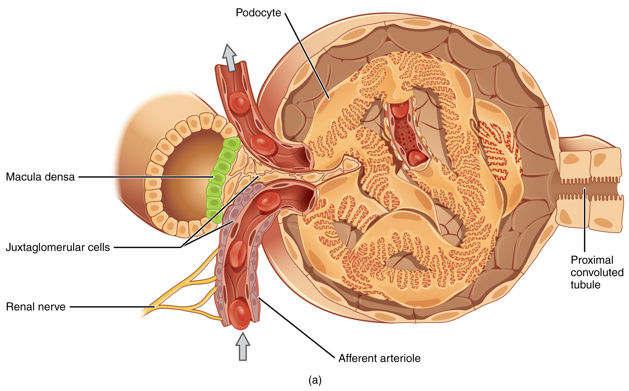
The renin–angiotensin–aldosterone system is the kidney’s main route for raising blood pressure. It begins with specialised cells in the wall of the afferent arteriole, part of the juxtaglomerular apparatus, which detect a fall in blood pressure or a fall in sodium delivery; sympathetic stimulation also triggers them. These cells release renin into the blood. Renin converts a liver protein called angiotensinogen into angiotensin I, which is inactive. As blood passes through the lungs, an enzyme called angiotensin-converting enzyme (ACE) converts angiotensin I into angiotensin II. Angiotensin II is the active hormone: it constricts blood vessels directly, which raises pressure; it stimulates aldosterone release, which retains sodium and water; it stimulates ADH release, which retains water; and it stimulates thirst, so more fluid is taken in. Together these actions restore blood volume and pressure.
For example: ACE inhibitors lower blood pressure because they block the conversion of angiotensin I to angiotensin II, reducing vasoconstriction and aldosterone release.
 Enlarge illustration ↗
Find where the tubule returns beside the glomerular arterioles. This close relationship links information from tubular fluid with control of blood flow and renin release. The macula densa and juxtaglomerular cells have different sensing and signalling roles.
Image source and licence
OpenStax Anatomy and Physiology · Source image · CC BY 4.0. Original image, reproduced without alteration.
Opposing signals and clinical links
Blood pressure is not controlled by one pathway alone. When blood volume rises, stretch receptors in the atria of the heart release atrial natriuretic peptide (ANP), and the ventricles release a similar hormone, BNP. These hormones promote sodium and water excretion by the kidneys, dilate blood vessels and reduce renin release, so they lower volume and pressure. The sympathetic nervous system also acts on the kidney, constricting renal vessels and stimulating renin release when pressure falls. The balance between the renin–angiotensin–aldosterone system and natriuretic peptides determines whether sodium and water are retained or excreted. Many common drugs act on these pathways: ACE inhibitors and angiotensin receptor blockers reduce angiotensin II effects, aldosterone antagonists block aldosterone, and diuretics increase sodium and water excretion. BNP is also measured in clinical practice as a marker of heart failure.
For example: A patient with heart failure may have a raised BNP level, because stretched ventricles release the hormone as pressures rise.
Key facts to remember
- The kidneys regulate blood volume, and therefore long-term blood pressure, by adjusting sodium and water excretion.
- ADH from the posterior pituitary increases collecting-duct water permeability via aquaporins, so urine becomes more concentrated.
- Aldosterone from the adrenal cortex increases sodium reabsorption and potassium secretion in the distal nephron; water follows the sodium.
- Renin from juxtaglomerular cells starts the cascade: angiotensinogen → angiotensin I → (ACE) → angiotensin II, which constricts vessels and stimulates aldosterone, ADH and thirst.
- ANP and BNP from the heart promote sodium and water excretion and lower pressure; ACE inhibitors, angiotensin receptor blockers, aldosterone antagonists and diuretics act on these pathways.
Example · Follow the response to dehydration. Water loss raises blood osmolarity, and osmoreceptors trigger ADH release. ADH makes the collecting ducts permeable to water, so water is reclaimed and a small volume of concentrated urine is produced. If blood pressure also falls, juxtaglomerular cells release renin. Renin starts the cascade that produces angiotensin II, which constricts vessels, stimulates aldosterone to retain sodium and water, stimulates ADH and triggers thirst. Sodium and water are retained, blood volume rises and pressure returns towards normal. When volume is restored, the signals fade and ANP from stretched atria promotes sodium and water excretion.
Read full notes
Read one topic at a time. These notes explain the mechanisms and relationships behind the key facts.
Why volume and pressure need regulation
Blood pressure has to be high enough to perfuse the brain and kidneys when a person stands, yet low enough to avoid damaging vessels over decades. The body achieves this with two systems working on different timescales. The heart and blood vessels adjust within seconds by changing rate and diameter, which is useful for sudden changes such as standing up or starting to run. The kidneys adjust over hours and days by changing how much sodium and water leave the body, which is what determines the long-term level of pressure. Because sodium is the main solute that holds water in the extracellular fluid, regulating sodium is equivalent to regulating volume. This is why almost all chronic blood pressure problems have a renal component, and why drugs that increase sodium excretion are so widely used.
ADH and water reabsorption
ADH is a good example of a hormone whose effect is visible in a single measurement. When ADH is high, the collecting ducts are permeable to water and urine is concentrated; when ADH is low, they are impermeable and urine is dilute. The hormone does not create the gradient that draws water out, because that gradient is built by the loop of Henle; ADH simply controls whether the collecting duct can use it. This division of labour is important: it means the kidney must maintain both the medullary gradient and the hormonal signal, and failure of either causes a concentrating defect. ADH release is controlled by osmoreceptors, which are extremely sensitive, and the response is rapid, so urine concentration changes within minutes of a change in blood osmolarity.
Aldosterone and sodium reabsorption
Aldosterone is the body's main sodium-retaining hormone, and its effects are wider than blood volume alone. By increasing sodium reabsorption and potassium secretion in the distal nephron, it links sodium balance, potassium balance and blood pressure. It is released in response to angiotensin II, to high blood potassium and to low blood sodium, so it responds both to the circulation and to the electrolyte composition of the blood. Because water follows sodium, prolonged aldosterone action expands blood volume and raises pressure. This is why drugs that block aldosterone are useful in hypertension and heart failure, and why conditions that produce too much aldosterone cause sodium retention, potassium loss and high blood pressure together.
The renin–angiotensin–aldosterone system
The renin–angiotensin–aldosterone system is powerful because it acts on four fronts at once. Angiotensin II narrows blood vessels, which raises pressure immediately. It stimulates aldosterone, which retains sodium and water over hours. It stimulates ADH, which retains water. And it stimulates thirst, which increases intake. Because the system is triggered by the kidney's own sensors, it responds to what the kidney perceives as inadequate perfusion, which may occur even when measured blood pressure appears normal. The system is also the target of several drug classes, which is why understanding its steps is clinically valuable: blocking renin, ACE or the angiotensin II receptor all reduce its effects, though by different mechanisms and with different side effects.
Opposing signals and clinical links
The body does not only defend against low volume; it also has mechanisms for getting rid of excess. Natriuretic peptides released by stretched cardiac chambers promote sodium and water excretion, dilate vessels and suppress renin, so they act as a brake on the renin–angiotensin–aldosterone system. The two systems therefore form a balance that responds to volume status in both directions. Clinically, this balance explains the logic of treatment. Diuretics increase sodium and water loss. ACE inhibitors and angiotensin receptor blockers reduce angiotensin II effects. Aldosterone antagonists block the sodium-retaining hormone. Because all of these lower volume or resistance, they lower pressure, and their side effects also follow from the pathways they affect.
Explore the diagram
Start with the whole view, then select a numbered structure. Compare the additional illustrations to connect what a structure looks like with what it does.
A tubule exchanges with blood
Enlarged transport schematic, not one specific tubular segment. Follow arrowheads: reabsorption is toward blood, secretion toward urine. Different transporters act in different nephron regions.
Tubular fluid contains the substances that remain for excretion. Urine volume and composition change as transport occurs along the nephron.
Follow fluid through a nephron
One simplified nephron with its collecting duct; neighbouring nephrons are omitted. Blood and tubular fluid occupy separate spaces. The cortex is above the dashed boundary.
The glomerular capillaries lie inside a capsule. Filtration moves water and small solutes from blood into the capsular space; cells and most proteins remain in blood.
Connect the illustrations to the key ideas
ADH AND WATER · osmoreceptors → posterior pituitary → aquaporins
High ADH concentrates urine; low ADH produces dilute urine.
RAAS AND ALDOSTERONE · renin → angiotensin I → ACE → angiotensin II → aldosterone
Sodium and water are retained and vessels constrict, raising pressure.
OPPOSING SIGNALS · ANP and BNP promote sodium and water excretion
The balance between RAAS and natriuretic peptides sets volume and pressure.
Core Concepts Defined
- Osmoreceptor
- A receptor in the hypothalamus that detects changes in blood solute concentration.
- Antidiuretic hormone (ADH)
- A hormone from the posterior pituitary that increases water reabsorption in the collecting ducts.
- Aquaporin
- A water channel inserted into collecting-duct cells under the influence of ADH.
- Diuresis
- The production of a large volume of urine.
- Diabetes insipidus
- A condition in which ADH is absent or ineffective, causing large volumes of dilute urine.
- Aldosterone
- A mineralocorticoid from the adrenal cortex that increases sodium reabsorption and potassium secretion.
- Mineralocorticoid
- A hormone that regulates sodium and potassium balance, such as aldosterone.
- Renin
- An enzyme released by juxtaglomerular cells that starts the angiotensin cascade.
- Angiotensinogen
- A liver protein converted by renin into angiotensin I.
- Angiotensin I
- The inactive product of renin action, converted to angiotensin II by ACE.
- Angiotensin II
- The active hormone that constricts vessels and stimulates aldosterone, ADH and thirst.
- Angiotensin-converting enzyme (ACE)
- The enzyme, abundant in the lungs, that converts angiotensin I to angiotensin II.
- Juxtaglomerular apparatus
- The structure containing the cells that release renin and the macula densa that senses sodium.
- Macula densa
- A group of cells in the distal tubule that senses sodium chloride delivery and signals renin release.
- Atrial natriuretic peptide (ANP)
- A hormone from stretched atria that promotes sodium and water excretion and lowers pressure.
- ACE inhibitor
- A drug that blocks angiotensin II formation, lowering blood pressure.
- Diuretic
- A drug that increases sodium and water excretion by the kidneys.
Go deeper · worked example
The response to dehydration
- Water is lost. Sweating, breathing or reduced intake lowers blood volume and raises the concentration of solutes in the blood, so blood osmolarity rises.
- Osmoreceptors detect the change. Cells in the hypothalamus sense the higher osmolarity and trigger ADH release from the posterior pituitary.
- ADH acts on the collecting ducts. Aquaporins are inserted into the duct cells, water is drawn out into the salty medulla and reabsorbed, and a small volume of concentrated urine is produced.
- Falling pressure triggers renin. If blood pressure or sodium delivery falls, juxtaglomerular cells release renin, which converts angiotensinogen to angiotensin I and, through ACE in the lungs, to angiotensin II.
- The response is integrated. Angiotensin II constricts vessels and stimulates aldosterone, ADH and thirst. Sodium and water are retained, the person drinks, blood volume rises and pressure returns towards normal; when volume is restored, ANP from stretched atria promotes excretion of the excess.
Explain it yourself · Explain why a person who has lost a large volume of sweat produces a small volume of concentrated urine.
Reveal a model explanation
Sweat loss removes water and raises the concentration of solutes in the blood, so blood osmolarity increases. Osmoreceptors in the hypothalamus detect this and trigger ADH release from the posterior pituitary. ADH acts on the collecting ducts, inserting aquaporins that make them permeable to water. Because the medulla surrounding the ducts is salty, water is drawn out of the duct and reabsorbed into the blood, leaving a smaller volume of more concentrated urine. If blood pressure also falls, renin is released and the renin–angiotensin–aldosterone system adds vasoconstriction, sodium and water retention and thirst, which together restore blood volume and pressure.
Check
Answer three questions from this section — one easy, one medium and one hard. If you miss one, you get a fresh set with explanations for the last attempt.
Practise this sectionOpen all questions and filter by difficulty or Bloom levelReferences and review status
OpenStax, Anatomy and Physiology 2e, 25.8 Regulation of Urine Concentration, 25.9 Regulation of Fluid Volume and Composition and 26.3 Electrolyte Balance; Tortora & Derrickson, Principles of Anatomy and Physiology, 12e, ch. 26. Author review only; medical and French-language expert review pending.
Original teaching schematics. English and French wording is author-reviewed for study; anatomy and French-language expert review is pending. Checks are practice only, not a graded examination.
Practice progress is saved on this device for the current learner.