SECTION 4.1
Plasma and formed elements
Identify the plasma and the formed elements of blood, and state the main role of each component.
Learn
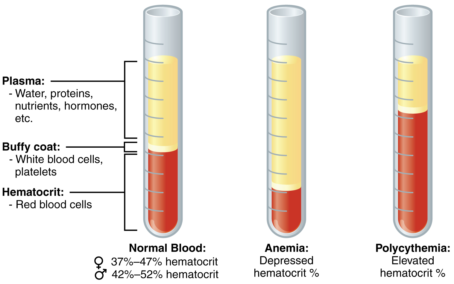
Blood is a connective tissue, but an unusual one: its matrix is a liquid. That liquid is plasma, and the cells and cell fragments suspended in it are called the formed elements. An adult has roughly five litres of blood, and every part of it performs a job. Red blood cells carry oxygen. White blood cells defend the body. Platelets help stop bleeding. Plasma carries the cells, dissolves nutrients and wastes, distributes heat and transports hormones and proteins. If a sample of blood is spun in a centrifuge, it separates into three visible layers: a large pale layer of plasma, a thin buffy coat containing white cells and platelets, and a red layer of packed red cells. Learning the composition of those layers gives you the vocabulary to interpret a blood test and to understand what happens when a component is too high or too low.
Key Concepts & Topics
Blood as a connective tissue
Blood is classified as connective tissue because it consists of cells scattered in a matrix, even though its matrix is liquid rather than solid or gel-like. The matrix is plasma, a pale yellow fluid that makes up a little more than half of blood volume. The cells and cell fragments are the formed elements, and they make up the rest. Blood has three broad functions. Transport: it carries oxygen from the lungs, nutrients from the gut, hormones from glands and waste products to the kidneys and lungs. Regulation: it distributes heat, helps control pH through buffers and influences water balance. Protection: white blood cells and plasma proteins defend against infection, and platelets and clotting proteins prevent blood loss. Recognising these three functions helps organise everything else you learn about blood.
For example: When you exercise, blood carries heat from the working muscles to the skin, where it is lost — a regulation function in action.
 Enlarge illustration ↗
Notice how much of the sample is liquid plasma. Red cells settle below it after centrifugation; white cells and platelets occupy a much thinner intermediate layer. The tube separates components for study—blood normally circulates as a mixture.
Image source and licence
OpenStax College · Source image · CC BY 3.0. Original image, reproduced without alteration.
Plasma and its proteins
Plasma is about 92% water, with dissolved substances making up the rest. It carries nutrients such as glucose, amino acids and lipids; ions such as sodium, potassium and calcium; waste products such as urea; gases in small amounts; and hormones. Its most distinctive solutes are the plasma proteins, which fall into three main groups. Albumins are the most abundant and are the main contributors to the osmotic pressure that holds water inside the vessels. Globulins include the antibodies that fight infection and the carrier proteins that transport lipids, hormones and iron. Fibrinogen is a clotting protein that is converted into fibrin when bleeding occurs. Plasma from which the clotting proteins have been removed is called serum, which is why a clotted blood sample yields serum rather than plasma. Because plasma proteins hold water in the vessels, losing them causes fluid to move into the tissues and produce oedema.
For example: In severe malnutrition or liver disease, albumin production falls, plasma osmotic pressure drops, and fluid leaks into the tissues, producing swelling.
Red blood cells
Red blood cells, also called erythrocytes, are the most numerous formed elements. A mature human red blood cell is a biconcave disc: thinner in the middle than at the edges. This shape gives a large surface area for exchanging gases and makes the cell flexible enough to pass through narrow capillaries. It has no nucleus and few organelles, which leaves more room for haemoglobin, the iron-containing protein that binds oxygen. Each red cell also carries antigens on its surface that determine blood group. A typical count is about five million cells per microlitre, and the cells live roughly 120 days before being removed by the spleen and liver. Their main job is to transport oxygen, and they also carry a share of the carbon dioxide produced by tissues.
For example: The biconcave shape is easy to remember from its consequence: a flat disc folds in the middle so it can bend as it squeezes through a capillary narrower than itself.
Common misconception: A mature human red blood cell has no nucleus, but it is not dead. It remains metabolically active, and it is a cell, not a fragment.
White blood cells
White blood cells, or leukocytes, defend the body. They are far fewer than red cells — normally about 5,000 to 10,000 per microlitre — and they contain a nucleus. They are grouped into five main types. Neutrophils are the most abundant and are the first responders to bacterial infection; they engulf microbes by phagocytosis. Eosinophils are involved in fighting parasites and in allergic responses. Basophils are the rarest; they release histamine and heparin and contribute to inflammation and allergic reactions. Lymphocytes include the T cells and B cells of immunity; B cells produce antibodies, and T cells help coordinate and carry out immune responses. Monocytes are the largest; they circulate, enter tissues and become macrophages that engulf debris and microbes. White cells can leave the bloodstream by squeezing between capillary cells, a process called diapedesis, so they can reach the site of infection.
For example: In acute appendicitis, the white cell count often rises because neutrophils are being mobilised to the inflamed tissue.
 Enlarge illustration ↗
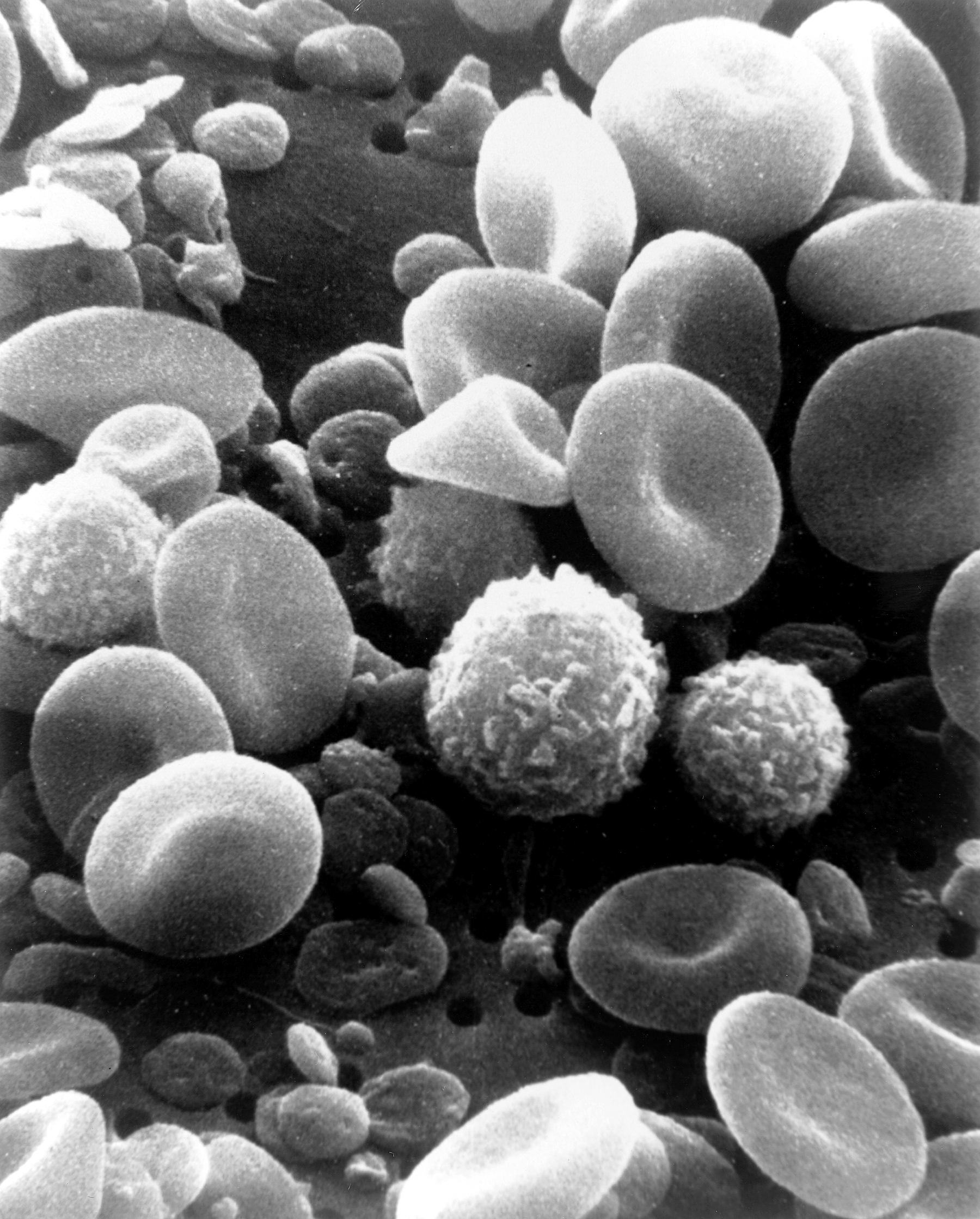
Look for the many disc-shaped red cells and the larger cells with uneven surfaces. This electron-microscope view reveals surface texture; it looks different from a stained blood film viewed with a light microscope.
Image source and licence
Bruce Wetzel (photographer). Harry Schaefer (photographer) · Source image · Public domain. Original image, reproduced without alteration.
Platelets
Platelets, also called thrombocytes, are not whole cells. They are small fragments pinched off from very large cells called megakaryocytes in the bone marrow. They have no nucleus and circulate for about a week to ten days. Their job is haemostasis: when a vessel is damaged, platelets stick to the exposed tissue, change shape, clump together to form a plug, and release substances that attract more platelets and help the clotting process. A normal platelet count is roughly 150,000 to 400,000 per microlitre. Too few platelets causes easy bruising and prolonged bleeding; too many increases the risk of unwanted clotting. Platelets are therefore essential for the first response to injury, even though they are not complete cells.
For example: When you cut yourself, the bleeding usually stops within minutes because platelets form a plug while clotting proteins build a firmer fibrin mesh.
Key facts to remember
- Blood is a connective tissue whose matrix is the fluid plasma and whose cells are the formed elements.
- Plasma carries nutrients, ions, wastes, gases and hormones, and contains albumins, globulins and fibrinogen.
- Red blood cells are biconcave, lack a nucleus when mature, are filled with haemoglobin and live about 120 days.
- The five white blood cell types are neutrophils, eosinophils, basophils, lymphocytes and monocytes.
- Platelets are cell fragments from megakaryocytes and form the first plug when a vessel is damaged.
Example · A blood sample is spun in a centrifuge. About 55% of the volume settles as pale plasma at the top. Below it is a thin buffy coat of white cells and platelets, usually less than 1% of the sample. The remaining 45% is packed red blood cells. The percentage of the sample occupied by red cells is the haematocrit. A low haematocrit suggests anaemia or fluid overload; a high one suggests dehydration or too many red cells. The buffy coat is thin because white cells and platelets are far fewer, but a change in its size or in the white cell count is an important clue to infection or immune problems.
Read full notes
Read one topic at a time. These notes explain the mechanisms and relationships behind the key facts.
Blood is a connective tissue
Calling blood a connective tissue surprises many learners because we usually picture connective tissue as solid: tendon, cartilage or bone. The defining feature of connective tissue, however, is not hardness but structure: cells scattered in a matrix. In blood the cells are the formed elements and the matrix is plasma, so the definition fits. This classification also explains blood’s functions. Like other connective tissues it supports and connects — it links every organ to every other by transporting substances between them — and like some connective tissues it stores and protects. Once you see blood as a connective tissue with a fluid matrix, its composition and its roles stop seeming arbitrary.
Plasma and its proteins
Plasma is mostly water, which is why it can dissolve and carry so many substances, but its proteins give it properties water alone does not have. Albumin, the most abundant, is too large to leave the capillaries easily, so it holds water inside the vessels by osmotic pressure; without it, fluid leaks into the tissues. Globulins include the antibodies that recognise specific microbes and the carrier proteins that ferry lipids, hormones and iron through the blood. Fibrinogen is kept ready for clotting and is converted into fibrin threads when a vessel is damaged. Because these proteins are made mainly in the liver, liver disease changes plasma composition and can cause bleeding problems, swelling or both. Plasma also carries the waste products that the kidneys remove, which is why kidney failure alters plasma chemistry.
Red blood cells and haemoglobin
The red blood cell is a study in efficiency. Losing its nucleus and most organelles frees space for haemoglobin, the protein that actually carries oxygen; each cell contains roughly 250 million haemoglobin molecules. The biconcave shape increases surface area for gas exchange and gives flexibility for narrow capillaries. The cell also carries surface antigens, notably the A and B antigens of the ABO system and the Rh antigen, which determine blood groups and matter greatly in transfusion. Because the cell cannot repair itself fully, it survives only about 120 days before being removed, mainly by the spleen and liver, and its iron is recycled. New red cells are produced in the bone marrow under the control of the hormone erythropoietin, released by the kidneys when oxygen delivery falls.
The five white blood cell types
White blood cells are the body’s mobile defence force, and each type has a specialised role. Neutrophils arrive first at a bacterial infection and are the most numerous; they are fast, short-lived phagocytes. Eosinophils are less numerous and specialise against parasites and in allergic reactions. Basophils are rare and release histamine and heparin, promoting inflammation and preventing unwanted clotting. Lymphocytes are the basis of specific immunity: B cells make antibodies that bind particular microbes, and T cells directly destroy infected cells or coordinate other immune cells. Monocytes circulate before entering tissues, where they enlarge into macrophages that clean up debris and microbes and also present information to lymphocytes. A differential white cell count reports the proportion of each type, and a shift in the proportion is a clue to the kind of problem.
Platelets and why they are not whole cells
Platelets are produced in the bone marrow by megakaryocytes, enormous cells that remain in the marrow while breaking off thousands of small membrane-bound fragments into the blood. Each fragment has no nucleus but contains granules of substances needed for clotting and for attracting other platelets. Their small size and large number make them ideal for patrolling: they circulate close to the vessel wall and can respond within seconds to a break. When they adhere to exposed tissue, they change shape, spread and aggregate into a plug. They also release factors that start the clotting cascade, studied in section 4.4. Because they are fragments rather than cells, they cannot divide, and their number depends on continuous production in the marrow and on removal by the spleen.
Explore the diagram
Start with the whole view, then select a numbered structure. Compare the additional illustrations to connect what a structure looks like with what it does.
The components of blood
A magnified teaching view. Cells and fragments are suspended in plasma; colours are illustrative.
More controls
01 / 4 Red blood cell
Red blood cell
The flattened disc has a thinner centre. Haemoglobin inside carries most blood oxygen.
Connect the illustrations to the key ideas
PLASMA · water, proteins, nutrients, wastes, hormones
The liquid matrix: it transports substances and its proteins hold water in the vessels.
FORMED ELEMENTS · red cells carry O₂; white cells defend
Erythrocytes are biconcave and haemoglobin-filled; five leukocyte types provide defence.
PLATELETS · cell fragments that form the first plug
They adhere, aggregate and release clotting factors within seconds of vessel injury.
Core Concepts Defined
- Blood
- A connective tissue of cells and cell fragments suspended in the fluid matrix plasma.
- Plasma
- The liquid matrix of blood, about 92% water, carrying dissolved substances and proteins.
- Serum
- Plasma from which the clotting proteins have been removed.
- Formed elements
- The cells and cell fragments of blood: red cells, white cells and platelets.
- Albumin
- The most abundant plasma protein; it holds water in the vessels by osmotic pressure.
- Globulin
- A plasma protein group that includes antibodies and carrier proteins.
- Fibrinogen
- A plasma clotting protein converted to fibrin during haemostasis.
- Erythrocyte (red blood cell)
- A biconcave, nucleus-free cell filled with haemoglobin that transports oxygen.
- Haemoglobin
- The iron-containing protein in red cells that binds oxygen.
- Leukocyte (white blood cell)
- A nucleated blood cell that defends the body; there are five main types.
- Neutrophil
- The most abundant white cell; a first responder that engulfs bacteria.
- Eosinophil
- A white cell involved in parasite defence and allergic responses.
- Basophil
- The rarest white cell; it releases histamine and heparin.
- Lymphocyte
- A white cell of immunity; B cells make antibodies and T cells coordinate responses.
- Monocyte
- The largest white cell; it becomes a macrophage in the tissues.
- Diapedesis
- The passage of white cells out of capillaries between the endothelial cells.
- Platelet (thrombocyte)
- A cell fragment from a megakaryocyte that forms the first plug after injury.
- Haemostasis
- The process that stops bleeding and keeps blood inside damaged vessels.
- Haematocrit
- The percentage of blood volume occupied by red blood cells.
- Buffy coat
- The thin layer of white cells and platelets seen after centrifuging blood.
Go deeper · worked example
Reading a spun blood sample
- Separate the sample. Centrifuging packs the denser cells at the bottom and leaves the lighter fluid on top, producing three layers.
- Identify the top layer. The pale yellow layer is plasma. It carries water, nutrients, wastes, hormones and the plasma proteins, so it is both a transport medium and a protein reservoir.
- Identify the middle layer. The thin buffy coat contains white blood cells and platelets. It is small because those elements are far less numerous than red cells, but it is the layer that becomes important in infection and immunity.
- Identify the bottom layer. The red layer is packed red blood cells, which carry oxygen bound to haemoglobin. The percentage of the sample they occupy is the haematocrit.
- Interpret the proportions. A low red layer suggests anaemia or dilution; a high one suggests dehydration or excess red cell production. A large buffy coat or a raised white cell count suggests infection or an immune disorder.
Explain it yourself · Explain what the haematocrit measures and why it does not measure white blood cells or platelets.
Reveal a model explanation
The haematocrit is the percentage of the blood sample occupied by packed red blood cells. It reflects red cell number and size in relation to plasma volume. White cells and platelets are far less numerous, and together they form only a thin buffy coat, usually under 1% of the sample. Their contribution to the volume is therefore too small to be measured this way; they are assessed by a white cell count and a platelet count instead. The haematocrit tells you about oxygen-carrying capacity and about the balance between cells and plasma.
Check
Answer three questions from this section — one easy, one medium and one hard. If you miss one, you get a fresh set with explanations for the last attempt.
Practise this sectionOpen all questions and filter by difficulty or Bloom levelReferences and review status
OpenStax, Anatomy and Physiology 2e, 18.1 Functions and Properties of Blood, 18.2 Production of the Formed Elements and 18.3 Erythrocytes; Tortora & Derrickson, Principles of Anatomy and Physiology, 12e, ch. 19. Author review only; medical and French-language expert review pending.
Original teaching schematics. English and French wording is author-reviewed for study; anatomy and French-language expert review is pending. Checks are practice only, not a graded examination.
Practice progress is saved on this device for the current learner.