SECTION 3.2
Muscle and nervous tissues
Distinguish the three muscle tissue types and nervous tissue, and relate each structure to how it produces movement or signals.
Learn
Two tissue families specialise in action. Muscle tissue produces movement by shortening: its cells are packed with contractile proteins that pull the cell shorter, and because the cells are anchored, that shortening moves something. Nervous tissue produces and conducts electrical signals: its cells detect changes, process information and send instructions to muscles and glands. Together they form the body’s movement and control systems. There are three kinds of muscle — skeletal, cardiac and smooth — and they differ in appearance, in whether we control them consciously, and in where they are found. Nervous tissue consists of neurons, which carry signals, and supporting cells, which protect, nourish and insulate them. Understanding their structure explains why a muscle can contract quickly and reliably and how a nerve can carry a signal long distances.
Key Concepts & Topics
Muscle tissue: built to shorten
Muscle cells contain long protein filaments, mainly actin and myosin, arranged so that they can slide past one another and pull the cell shorter. This shortening is contraction, and it is an active process that uses ATP. Because each muscle cell is attached at its ends to something — bone, another muscle cell or the wall of a hollow organ — contraction produces movement, maintains posture or moves material along a passage. Muscle tissue is also a major source of body heat, because contraction releases energy as heat. Three types exist. Skeletal muscle is attached to bone and produces body movement; cardiac muscle forms the heart wall and pumps blood; smooth muscle lines hollow organs and moves their contents. The three differ in appearance, in control and in endurance, but all share the same basic ability to shorten.
For example: When you lift a cup, the biceps shortens, pulling on a tendon attached to bone. Nothing is pushed; the movement comes from the muscle getting shorter.
Skeletal muscle
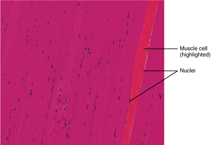
Skeletal muscle is the type you can see and usually think of first. Its cells are long, cylindrical and have many nuclei, and under the microscope they show alternating light and dark bands, so they are described as striated. Skeletal muscle is voluntary: you decide when to move it, though it also takes part in reflexes and in maintaining posture. It contracts quickly and powerfully but tires when it works hard for a long time. Each muscle contains bundles of fibres, and each fibre receives a nerve ending, so the nervous system can activate the muscle in graded steps: a few fibres for a light task, many for a strong one. This precise control is what allows fine movements such as writing and powerful movements such as jumping.
For example: Writing uses small, precise contractions in the hand, while jumping uses many fibres at once in the legs — the same tissue, activated differently.
 Enlarge illustration ↗
This is real tissue under a microscope. Look for parallel fibres and the dark nuclei. A single skeletal muscle fibre contains many nuclei; the staining makes structures visible and does not represent their natural colour.
Image source and licence
OpenStax · Source image · CC BY 4.0. Original image, reproduced without alteration.
Cardiac and smooth muscle
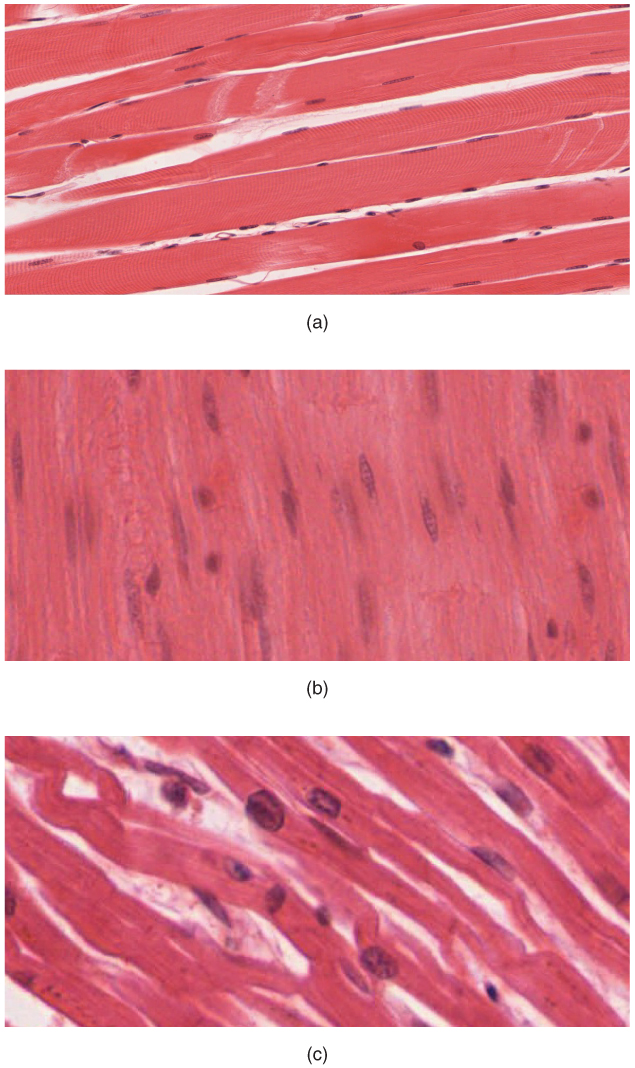
Cardiac muscle forms the wall of the heart. Its cells are striated like skeletal muscle but are shorter, branched and joined end to end by special junctions called intercalated discs, which let the electrical signal spread quickly from cell to cell so the heart contracts as a unit. Cardiac muscle is involuntary: we do not consciously control the heartbeat, and the tissue has its own rhythm, modified by nerves and hormones. It is highly resistant to fatigue because the heart must work continuously for a lifetime. Smooth muscle is found in the walls of hollow organs such as the stomach, intestines, bladder, uterus and blood vessels. Its cells are spindle-shaped with a single nucleus and have no visible striations. Smooth muscle is also involuntary; it contracts slowly and can sustain contraction for long periods, moving food, urine or blood, and it can stretch and still work, which is essential in a bladder or a uterus.
For example: Your stomach churns food and your arteries adjust their width without any conscious decision, because both contain smooth muscle.
Common misconception: Involuntary does not mean uncontrolled. Cardiac and smooth muscle are regulated by nerves, hormones and local signals; they simply are not directed by conscious will.
 Enlarge illustration ↗
Panels (a), (b) and (c) show skeletal, smooth and cardiac muscle respectively. Compare the tissue photographs with the simplified Explore drawings. Skeletal and cardiac muscle show striations; smooth muscle does not. Use both cell shape and the arrangement of nuclei when comparing them.
Image source and licence
OpenStax College · Source image · CC BY 4.0. Original image, reproduced without alteration.
Nervous tissue: cells that signal
Nervous tissue is built to generate and conduct electrical signals quickly. It contains two broad cell groups. Neurons are the signalling cells; they have a cell body containing the nucleus, dendrites that receive signals from other cells, and usually a single long axon that carries the signal away to its target. Some axons are wrapped in a fatty insulating layer called myelin, which speeds conduction. Neuroglia, or glial cells, are the supporting cells: they protect and nourish neurons, remove waste and debris, form myelin and help maintain the environment around the neurons. Nervous tissue is found in the brain, spinal cord and nerves, and it controls and coordinates almost every other tissue by sending rapid instructions and receiving feedback. Unlike epithelium, nervous tissue has very little matrix, but it is not a simple sheet: its cells are organised into circuits.
For example: When you touch something hot, sensory neurons carry the signal to the spinal cord, where a circuit triggers motor neurons that make skeletal muscle pull your hand away.
Movement and control together
Muscle and nervous tissue depend on each other. Muscle contraction is triggered by signals from motor neurons; without them, even healthy muscle cannot act. Nervous tissue, in turn, depends on muscle for output: it decides what movement should happen, but the muscle carries it out. Both depend on ion gradients and ATP, which is why conditions that disturb ion balance or energy supply affect movement and signalling early. The two families also share an electrical mechanism based on the movement of ions across cell membranes, studied in sections 2.2 and 2.3. Recognising this shared basis helps explain why disorders of electrolytes can cause both muscle weakness and abnormal nerve function at the same time, and why fatigue has effects on both systems.
For example: A low potassium level can cause both muscle weakness and disturbed heart rhythm, because muscle and nervous tissues both rely on ion gradients.
Key facts to remember
- Muscle tissue produces movement by shortening, using actin and myosin filaments and ATP.
- Skeletal muscle is striated and voluntary; cardiac muscle is striated, involuntary and joined by intercalated discs; smooth muscle is non-striated and involuntary.
- Cardiac muscle contracts as a unit and resists fatigue; smooth muscle contracts slowly, sustains contraction and can work while stretched.
- Nervous tissue contains neurons, which conduct signals, and neuroglia, which support, nourish and insulate them.
- Both muscle and nervous tissue depend on ion gradients and ATP, linking electrolyte and energy problems to movement and signalling.
Example · Standing up from a chair uses all three muscle types and nervous tissue at once. The brain sends signals along motor neurons to skeletal muscle in the legs, which shortens and lifts the body. Cardiac muscle keeps pumping faster to deliver more oxygen. Smooth muscle in the arteries adjusts their width to redirect blood, and smooth muscle in the gut continues moving its contents. Nervous tissue senses the change in posture and adjusts the signals continuously so you do not fall.
Read full notes
Read one topic at a time. These notes explain the mechanisms and relationships behind the key facts.
Built to shorten: the contractile cell
A muscle cell converts chemical energy into mechanical work. Its cytoplasm is filled with myofibrils, long bundles of actin and myosin filaments arranged in a repeating pattern. When a signal arrives, the filaments slide past one another, pulling the attachment points closer together, and the cell shortens. This needs ATP both to power the sliding and to reset the system afterwards. Because the filaments can only pull, muscles usually work in opposing pairs: one shortens to bend a joint, the other to straighten it. The striations visible under the microscope come from the regular arrangement of these filaments, which is why striated muscles contract quickly and powerfully, while smooth muscle, with its less ordered filaments, contracts more slowly but can sustain the effort.
The three muscle types compared
Skeletal muscle is built for speed and power under voluntary control. Its fibres are long and multinucleated, and each is activated by a motor neuron, so the nervous system can recruit exactly as many fibres as a task needs. Cardiac muscle is built for endurance and rhythmicity. Its cells are branched and linked by intercalated discs, allowing the electrical signal to pass freely so the heart contracts as one unit, and its own pacemaker tissue keeps it beating without conscious input. Smooth muscle is built for sustained, economical work in hollow organs. Its cells are spindle-shaped and arranged in sheets or layers, and its contraction moves contents, controls the width of passages and can continue for hours with little fatigue. The differences in form match exact differences in job.
Nervous tissue: cells that signal
Nervous tissue works by changing the electrical charge across neuron membranes. At rest, a neuron maintains a difference in charge between the inside and the outside, built by ion pumps and channels. A stimulus opens channels, ions move, and the charge reverses briefly; this reversal travels along the axon as a signal. At the end of the axon, the signal triggers the release of chemical messengers that carry the information across a small gap to the next cell. Myelin speeds the signal by allowing it to jump between gaps in the insulation. This system is fast, precise and adaptable, and because it depends on ion gradients, it is sensitive to anything that disturbs those gradients, such as changes in potassium, sodium or calcium.
Neurons and supporting cells
Neurons are often described as the wiring of the body, but they could not work alone. Neuroglia outnumber neurons and perform essential support tasks: some wrap axons in myelin to speed conduction, some remove debris and excess signalling molecules, some form the barrier that controls what reaches the nervous system from the blood, and others provide structural support and nutrients. Without this support, neurons lose their environment and fail. This division of labour explains why many neurological diseases involve supporting cells as much as neurons. It is also a good example of a general principle in this course: a tissue’s function depends not only on its main cells but on the cells and material around them.
Movement and control are one system
It is convenient to study muscle and nervous tissue separately, but they act as one system. A motor neuron decides when and how strongly a muscle contracts; a sensory neuron reports what happened, allowing the brain to adjust the next command. This loop explains the precision of human movement and why damage at any point disrupts it: injured nervous tissue prevents muscles from receiving instructions, and injured muscle prevents the nervous system from carrying them out. Both tissues also rely on the same ion gradients and ATP, so a single disturbance, such as a severe electrolyte imbalance, can produce weakness, abnormal sensation and disturbed heart rhythm together. Thinking of movement and control as a partnership is a better preparation for clinical practice than memorising them as separate lists.
Explore the diagram
Start with the whole view, then select a numbered structure. Compare the additional illustrations to connect what a structure looks like with what it does.
A nerve pathway and a neuron
The body view locates the nervous system; the enlarged neuron explains how a cell receives and passes on a signal.
More controls
01 / 7 Brain
Brain
The brain is part of the central nervous system and integrates information.
Illustration credits
- Mariana Ruiz Villarreal (LadyofHats) · Complete neuron cell diagram en.svg · Public domain.

Adapted for this lesson: labels removed, artwork cropped or recoloured, and interactive teaching overlays added. Adapted artwork retains its source licence.
Recognise the three muscle tissues
Enlarged longitudinal views, independently scaled. Compare stripes, branching and nuclear position before reading the labels.
Long, unbranched, striated fibres contain many peripheral nuclei. They produce voluntary movements such as lifting the forearm.
Connect the illustrations to the key ideas
SKELETAL MUSCLE · striated, voluntary, attached to bone
Contracts quickly and powerfully to move the body and maintain posture.
CARDIAC AND SMOOTH MUSCLE · involuntary: heart wall and hollow organs
Cardiac muscle pumps without fatigue; smooth muscle moves contents and adjusts vessel width.
NERVOUS TISSUE · neurons signal, neuroglia support
Electrical signals travel along axons and trigger chemical messages at the next cell.
Core Concepts Defined
- Muscle tissue
- Tissue specialised to contract and produce movement, heat and the movement of materials.
- Actin and myosin
- The contractile protein filaments that slide past one another to shorten a muscle cell.
- Contraction
- The active shortening of a muscle cell, which requires ATP.
- Skeletal muscle
- Striated, voluntary muscle attached to bone that produces body movement.
- Striated
- Showing alternating light and dark bands under the microscope.
- Voluntary
- Under conscious control.
- Involuntary
- Not under conscious control, though still regulated by nerves and hormones.
- Cardiac muscle
- Striated, involuntary muscle of the heart wall, joined by intercalated discs and resistant to fatigue.
- Intercalated disc
- A junction between cardiac muscle cells that lets the electrical signal spread rapidly.
- Smooth muscle
- Non-striated, involuntary muscle in the walls of hollow organs and vessels.
- Nervous tissue
- Tissue that generates and conducts electrical signals, made of neurons and neuroglia.
- Neuron
- The signalling cell of nervous tissue, with a cell body, dendrites and usually one axon.
- Dendrite
- A neuron extension that receives signals from other cells.
- Axon
- The long neuron extension that carries signals away towards a target.
- Neuroglia (glial cells)
- Supporting cells that protect, nourish, insulate and maintain the environment of neurons.
- Myelin
- A fatty insulating wrap around some axons that speeds signal conduction.
- Electrolyte
- A dissolved ion, such as sodium or potassium, that carries charge in body fluids.
Go deeper · worked example
One action, three muscle types and a nerve circuit
- Decide and send the signal. The brain generates a signal that travels along motor neurons to the skeletal muscles of the legs.
- Contract skeletal muscle. The signal triggers the muscle fibres to slide their actin and myosin filaments, shortening the cells, which pull on tendons and move the bones.
- Support the effort with cardiac muscle. As demand rises, the heart contracts faster and more strongly, joined by intercalated discs so its chambers squeeze as a unit.
- Redirect blood with smooth muscle. Smooth muscle in the arteries adjusts vessel width so more blood reaches the working muscles and less goes to resting tissue.
- Maintain balance with feedback. Sensory neurons report the change in posture, and the nervous system adjusts the motor signals continuously until the movement is complete and stable.
Explain it yourself · Explain why the same movement needs both nervous tissue and all three types of muscle.
Reveal a model explanation
Nervous tissue decides and directs: it generates the signal, carries it to the muscles and adjusts it using feedback, without which movement would be uncoordinated or impossible. Skeletal muscle produces the visible movement by shortening. Cardiac muscle supplies the extra blood flow the working muscles need. Smooth muscle redistributes that blood by changing vessel width and keeps internal organs working. Each tissue does one part of a single coordinated action, which is why a problem in any one of them changes the whole movement.
Check
Answer three questions from this section — one easy, one medium and one hard. If you miss one, you get a fresh set with explanations for the last attempt.
Practise this sectionOpen all questions and filter by difficulty or Bloom levelReferences and review status
OpenStax, Anatomy and Physiology 2e, 4.4 Muscle Tissue and 4.5 Nervous Tissue; Tortora & Derrickson, Principles of Anatomy and Physiology, 12e, ch. 4. Author review only; medical and French-language expert review pending.
Original teaching schematics. English and French wording is author-reviewed for study; anatomy and French-language expert review is pending. Checks are practice only, not a graded examination.
Practice progress is saved on this device for the current learner.