SECTION 8.5
Micturition and normal urine
Describe micturition and the normal physical and chemical characteristics of urine.
Learn
Urine is produced continuously, but it leaves the body intermittently. The process of emptying the bladder is called micturition, and it depends on a muscular sac, two sphincters and a reflex that can be overridden by conscious control. As the bladder fills, stretch receptors signal the spinal cord, which triggers contraction of the detrusor muscle and relaxation of the internal sphincter. In infants this reflex empties the bladder automatically. In older children and adults, higher centres can keep the external sphincter closed until it is convenient to void. Once urine is produced, its physical and chemical characteristics are informative: normal urine is pale yellow, clear, slightly acidic and contains mainly water, urea, creatinine and ions. This section describes how the bladder empties and what normal urine looks like.
Key Concepts & Topics
The bladder and its muscles
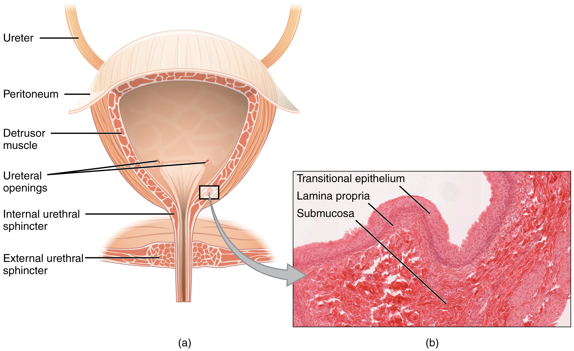
The urinary bladder is a hollow muscular organ in the pelvis. Its wall contains the detrusor muscle, a layer of smooth muscle that contracts to expel urine. The inner surface of the base has a smooth triangular area called the trigone, marked by the two openings of the ureters and the internal urethral opening. Two sphincters control the exit. The internal urethral sphincter is a thickening of smooth muscle at the bladder neck; it is involuntary and normally closed. The external urethral sphincter is made of skeletal muscle around the urethra and is under voluntary control. Because the bladder is a muscular sac with a stretchable lining, it can store several hundred millilitres of urine without a large rise in pressure, which allows urine to be held comfortably between voidings.
For example: Bladder capacity is roughly 400–600 mL in an adult, though the first urge to void is usually felt well before that volume.
 Enlarge illustration ↗
Trace urine from the two ureter openings into the bladder, then toward the urethra below. The detrusor is the muscular wall that squeezes during emptying. Compare the folded lining in the drawing with the tissue photograph.
Image source and licence
OpenStax College · Source image · CC BY 3.0. Original image, reproduced without alteration.
The micturition reflex
Micturition begins with stretch. As the bladder fills, stretch receptors in its wall send signals along pelvic nerve afferents to the sacral segments of the spinal cord. When the signal is strong enough, the spinal cord produces a reflex: parasympathetic fibres in the pelvic nerves stimulate the detrusor muscle to contract and relax the internal urethral sphincter. If the external sphincter is also relaxed, urine flows. As urine passes through the urethra, it stimulates receptors that reinforce the contraction, so the bladder empties completely. This spinal reflex is the whole mechanism in infants and in people with spinal cord injury above the sacral level, which is why emptying can occur without conscious control in those situations.
For example: A newborn voids by reflex alone, because the pathways for voluntary control have not yet matured.
Voluntary control of micturition
In older children and adults, the simple reflex is modified by higher centres. The pontine micturition centre coordinates the switch between storage and voiding, and the cerebral cortex can consciously inhibit the reflex by keeping the external urethral sphincter contracted. This is how a person can postpone voiding even when the bladder is full: the detrusor may contract briefly, but the external sphincter holds, and the urge subsides. When it is convenient, the cortex withdraws its inhibition, the external sphincter relaxes voluntarily, and the reflex completes the emptying. Continence is therefore a learned skill that depends on intact spinal pathways, an intact pontine centre and functioning cortical control, and it can be lost if any of these are damaged.
For example: After a spinal cord injury above the sacral segments, the reflex may still empty the bladder, but voluntary control is lost.
Normal urine: physical characteristics
Normal urine has a characteristic appearance. Volume is usually about one to two litres a day in an adult, depending on fluid intake, diet, activity and hormones; the kidneys must excrete at least about 500 millilitres a day to clear waste products. Colour is pale yellow to amber, produced mainly by urobilin, a pigment formed from the breakdown of haemoglobin. Freshly passed urine is usually clear, though it may become cloudy on standing as salts precipitate. It has a mild characteristic odour that becomes stronger and more ammonia-like as bacteria act on it. pH varies from about 4.5 to 8, being usually slightly acidic, and specific gravity, which reflects the concentration of dissolved solutes, ranges from about 1.001 when dilute to 1.035 when concentrated.
For example: A first-morning urine sample is usually more concentrated, with a higher specific gravity, because no fluid has been taken overnight.
Normal urine: chemical composition and abnormal findings
About 95% of urine is water; the remaining solutes include urea, the main nitrogenous waste, creatinine from muscle metabolism, uric acid, ammonia, and ions such as sodium, potassium, chloride, sulfate and phosphate. Small amounts of urobilinogen and hormones are also present. Normal urine contains no detectable glucose, protein, blood or ketones, so their presence is significant. Glucose indicates that the reabsorptive transport maximum was exceeded, as in diabetes. Protein suggests damage to the filtration barrier. Blood may indicate infection, stones or injury. Ketones indicate rapid fat breakdown, as in uncontrolled diabetes or prolonged fasting. Changes in volume also matter: polyuria is excessive urine output, oliguria is reduced output and anuria is almost none, and each points to different problems in filtration, reabsorption or drainage.
For example: A urine sample positive for blood and protein in a patient with flank pain may suggest a kidney stone or infection.
Key facts to remember
- Micturition is the emptying of the bladder, driven by contraction of the detrusor muscle and relaxation of the sphincters.
- The internal urethral sphincter is involuntary smooth muscle; the external urethral sphincter is voluntary skeletal muscle.
- Stretch receptors signal the sacral spinal cord, which triggers the micturition reflex through parasympathetic pelvic nerves.
- Higher centres, including the pontine micturition centre and the cerebral cortex, allow voiding to be postponed by keeping the external sphincter contracted.
- Normal urine is pale yellow, clear, usually slightly acidic (pH 4.5–8) with specific gravity 1.001–1.035, and contains water, urea, creatinine, ions and urobilin but no glucose, protein, blood or ketones.
Example · Follow a normal void. Urine drains from the collecting ducts into the calyces and pelvis, then down the ureters to the bladder by peristalsis. The bladder fills and its wall stretches; stretch receptors send signals to the sacral cord. The detrusor contracts and the internal sphincter relaxes through the pelvic nerves. If the external sphincter is relaxed voluntarily, urine flows through the urethra, and its passage reinforces the contraction until the bladder is empty. The process is automatic in infants and consciously controlled in adults.
Read full notes
Read one topic at a time. These notes explain the mechanisms and relationships behind the key facts.
The bladder and its muscles
The bladder is built for storage as well as emptying, and its structure reflects both jobs. The detrusor provides the force to expel urine; the trigone provides a smooth, fixed area where the ureters enter, so that urine does not reflux upward as the bladder contracts; and the two sphincters allow the exit to be closed or opened. The internal sphincter is smooth muscle and therefore involuntary, while the external sphincter is skeletal muscle and voluntary. This division is the anatomical basis of continence: the reflex controls the internal sphincter, and conscious control acts on the external one. The bladder's ability to expand without a large pressure rise is due to the folded lining and the elastic properties of the detrusor, and it is what allows comfortable storage between voidings.
The micturition reflex
The micturition reflex is a spinal cord reflex, which is why it works without the brain. Stretch receptors fire as the bladder fills, and when their input reaches a threshold the sacral cord switches the bladder from storage to voiding mode. The same reflex includes a positive feedback element: as urine enters the urethra, it stimulates receptors that increase detrusor contraction, so emptying continues once started. This is efficient, but it also means that the reflex alone cannot hold urine when it is triggered. In health, higher centres suppress the reflex during filling and permit it at a suitable time, which is why people can sleep through the night without voiding despite continuous urine production.
Voluntary control of micturition
Continence depends on three levels working together. The peripheral reflex provides the basic emptying mechanism; the pontine centre organises it into alternating storage and voiding phases; and the cerebral cortex provides voluntary control through the external sphincter. Damage at any level produces a different pattern. Loss of cortical control may cause urgency or incontinence; damage to the sacral cord or its nerves may cause retention with overflow; and damage above the sacral level may leave reflex emptying without voluntary control. Recognising these patterns is clinically useful because it localises the problem to a level of the nervous system, and it explains why continence is described as a learned skill rather than a simple reflex.
Normal urine: physical characteristics
The physical characteristics of urine are a rapid summary of kidney function and body state. Volume reflects fluid balance and hormonal control; colour reflects the concentration of pigments and, when abnormal, the presence of blood, bile or drugs; clarity reflects the presence of cells, crystals or fat; and specific gravity reflects how concentrated the urine is. Because these features are easy to measure, they are used routinely. A pale, dilute urine with low specific gravity after drinking a lot of water is normal, as is a darker, more concentrated urine in the morning. What matters is the pattern: persistent dilute urine suggests that ADH is absent or ineffective, while persistent concentrated urine may suggest dehydration or another cause of water conservation.
Chemical composition and abnormal findings
The chemical composition of urine tells the second half of the story. Water is the solvent, urea is the main nitrogenous waste from protein breakdown, creatinine comes from muscle metabolism at a steady rate and is used to estimate kidney function, and uric acid comes from nucleic acid breakdown. The various ions reflect dietary intake and the body's electrolyte balance. Because normal urine contains no glucose, protein, blood or ketones, each of these findings has a short differential diagnosis, which is why urine testing is such an efficient first investigation. Interpreting it requires linking the finding to the physiology: glucose to the transport maximum, protein to the filtration barrier, blood to injury or infection, and ketones to fat metabolism.
Explore the diagram
Start with the whole view, then select a numbered structure. Compare the additional illustrations to connect what a structure looks like with what it does.
Kidneys to bladder
Two kidneys drain through two ureters into one bladder. The urethra is the outlet.
More controls
01 / 6 Right kidney
Right kidney
The kidneys filter plasma and adjust water, salts and acid–base balance. The right often lies slightly lower.
Illustration credits
- Jmarchn · Urinary system.svg · CC BY-SA 3.0.

Adapted for this lesson: labels removed, artwork cropped or recoloured, and interactive teaching overlays added. Adapted artwork retains its source licence.
Storage and the bladder outlet
Simplified frontal section. Ureters enter the bladder; the urethra leaves it. The outlet arrangement varies with sex, so this is a functional schematic.
Paired tubes carry urine from the kidneys into the bladder. They are different from the single urethra, which carries urine out of the body.
Connect the illustrations to the key ideas
BLADDER FILLING · detrusor relaxes; stretch receptors signal
The internal sphincter stays closed and pressure rises slowly.
THE REFLEX · sacral cord → pelvic nerves → detrusor contracts
The internal sphincter relaxes; urethral flow reinforces the contraction.
VOLUNTARY CONTROL · pontine centre and cortex control the external sphincter
Voiding can be postponed until convenient; continence is learned.
Core Concepts Defined
- Micturition
- The process of emptying the urinary bladder.
- Detrusor muscle
- The smooth muscle of the bladder wall that contracts to expel urine.
- Trigone
- The smooth triangular area of the bladder base between the ureter openings and the urethra.
- Internal urethral sphincter
- The involuntary smooth muscle sphincter at the bladder neck.
- External urethral sphincter
- The voluntary skeletal muscle sphincter around the urethra.
- Pelvic nerve
- The nerve carrying parasympathetic fibres that trigger detrusor contraction.
- Pontine micturition centre
- The brainstem centre that coordinates storage and voiding of urine.
- Continence
- The learned ability to control when urine is voided.
- Incontinence
- Involuntary loss of urine.
- Urinary retention
- The inability to empty the bladder despite the urge to void.
- Specific gravity
- A measure of the concentration of dissolved solutes in urine.
- Urobilin
- The pigment formed from haemoglobin breakdown that gives urine its yellow colour.
- Polyuria
- Abnormally large urine output.
- Oliguria
- Abnormally reduced urine output.
- Anuria
- Absence of urine output.
- Glycosuria
- Glucose in the urine, usually from blood glucose above the renal threshold.
- Proteinuria
- Protein in the urine, suggesting damage to the filtration barrier.
- Haematuria
- Blood in the urine.
Go deeper · worked example
Following a normal void
- Urine reaches the bladder. Urine flows from the collecting ducts into the calyces and renal pelvis, then down the ureters by peristalsis into the bladder.
- The bladder fills and stretches. The detrusor relaxes and the wall expands, so pressure rises only slowly; stretch receptors begin to signal the sacral spinal cord.
- The reflex is triggered. When the signal is strong enough, parasympathetic fibres in the pelvic nerves cause the detrusor to contract and the internal sphincter to relax.
- Voluntary control decides the moment. The pontine centre and cerebral cortex can keep the external sphincter contracted, postponing voiding; when it suits, the cortex withdraws inhibition and the external sphincter relaxes.
- Emptying is completed. Urine flowing through the urethra stimulates receptors that reinforce the contraction, so the bladder empties fully and the cycle begins again as urine continues to arrive.
Explain it yourself · Explain why an infant voids automatically while an adult can choose when to void.
Reveal a model explanation
Bladder emptying is controlled by a spinal reflex. Stretch receptors in the bladder wall signal the sacral spinal cord, and parasympathetic fibres in the pelvic nerves cause the detrusor to contract and the internal sphincter to relax. In an infant this reflex operates on its own, because the pathways for voluntary control have not yet matured. In an adult the same reflex exists, but it is modified by higher centres: the pontine micturition centre coordinates storage and voiding, and the cerebral cortex can keep the external urethral sphincter contracted, holding urine in even when the detrusor contracts. When it is convenient, the cortex withdraws that inhibition, the external sphincter relaxes voluntarily, and the reflex completes emptying. Continence is therefore a learned control over an underlying reflex.
Check
Answer three questions from this section — one easy, one medium and one hard. If you miss one, you get a fresh set with explanations for the last attempt.
Practise this sectionOpen all questions and filter by difficulty or Bloom levelReferences and review status
OpenStax, Anatomy and Physiology 2e, 25.1 Physical Characteristics of Urine and 25.10 Micturition; Tortora & Derrickson, Principles of Anatomy and Physiology, 12e, ch. 26. Author review only; medical and French-language expert review pending.
Original teaching schematics. English and French wording is author-reviewed for study; anatomy and French-language expert review is pending. Checks are practice only, not a graded examination.
Practice progress is saved on this device for the current learner.