SECTION 7.2
Lymphatic vessels and lymph return
Trace lymph from a lymphatic capillary through vessels, nodes, trunks and ducts back to venous blood.
Learn
Once lymph has entered a lymphatic capillary, it must travel all the way back to the blood, and there is no heart to push it. The journey passes through a graded system of vessels: capillaries join to form collecting vessels, collecting vessels join to form trunks, and the trunks empty into two large ducts that return lymph to the veins at the base of the neck. Along the way, lymph passes through lymph nodes, which filter it and give immune cells a chance to inspect it. Because there is no central pump, lymph is moved by the squeezing of skeletal muscles, by breathing movements and by the contraction of smooth muscle in the vessel walls, with valves ensuring that it moves only forward. This section follows the route from capillary to vein and explains the forces that keep lymph moving.
Key Concepts & Topics
From capillary to collecting vessel
Lymphatic capillaries merge into larger collecting vessels. As the vessels get larger, their walls acquire more layers and more smooth muscle, and they come to resemble veins. Collecting vessels contain many valves, spaced closely together, which prevent lymph from flowing backward and help divide the column of lymph into short segments that can be squeezed forward. Along their course, collecting vessels pass through lymph nodes, often several in a row, and lymph must traverse the node before continuing. Afferent vessels bring lymph into a node; efferent vessels carry it out. Because lymph flows through nodes rather than bypassing them, the nodes act as filters and as surveillance points, and this is why an infection or a cancer spreading through lymph often affects the nodes first.
For example: A scratch on the hand may cause tenderness in the armpit days later, because lymph from the hand passes through the axillary nodes on its way back to the blood.
The lymphatic trunks
Collecting vessels converge into larger lymph trunks, each draining a region of the body. The lumbar trunks drain the lower limbs, the pelvis and the kidneys. The intestinal trunk drains the stomach, intestines, pancreas and spleen, and carries the fat-rich lymph called chyle after a meal. The bronchomediastinal trunks drain the thoracic wall, lungs and heart. The subclavian trunks drain the upper limbs, and the jugular trunks drain the head and neck. The lumbar and intestinal trunks commonly join to form a dilated sac called the cisterna chyli in the upper abdomen, which continues upward as the thoracic duct. The trunks are the collecting points of the lymphatic system, and knowing which trunk drains which region is the basis for understanding where cancer cells from a particular organ may travel.
For example: The intestinal trunk carries chyle, the milky fat-rich lymph, to the cisterna chyli and then up the thoracic duct.
The two ducts and what they drain
All lymph eventually enters one of two ducts. The thoracic duct is the larger. It begins at the cisterna chyli, ascends through the thorax and empties into the venous blood at the left venous angle, where the left subclavian and left internal jugular veins meet. It drains the entire body below the diaphragm and the left side above the diaphragm: both legs, the abdomen, the left arm and the left side of the head, neck and chest. The right lymphatic duct is much shorter. It drains only the right side above the diaphragm — the right arm and the right side of the head, neck and chest — and empties at the right venous angle. This asymmetry explains why a blockage of the thoracic duct causes widespread swelling, while a blockage of the right lymphatic duct affects only the upper right quadrant of the body.
For example: Lymph from the left foot, the abdomen and the left hand all return through the thoracic duct; lymph from the right hand returns through the right lymphatic duct.
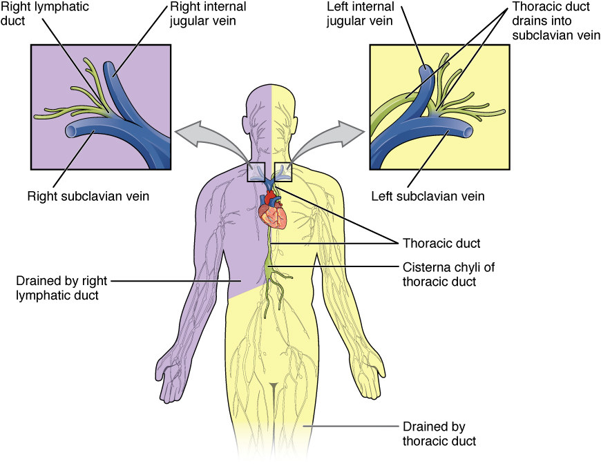 Enlarge illustration ↗
Use the patient’s right and left, not the viewer’s. Compare the small territory drained by the right lymphatic duct with the much larger territory of the thoracic duct. Both return lymph to veins near the base of the neck.
Image source and licence
OpenStax College · Source image · CC BY 3.0. Original image, reproduced without alteration.
How lymph is propelled
Lymph moves at low pressure and there is no central pump, so several mechanisms work together. Contraction of skeletal muscles during movement squeezes the collecting vessels, and valves ensure that the squeezed lymph moves forward rather than back. Breathing movements change pressure in the chest and abdomen, drawing lymph upward as you inhale. Pulsation of nearby arteries also compresses lymphatic vessels. Finally, the smooth muscle in the walls of larger lymphatic vessels contracts rhythmically on its own, and this contraction is increased by physical activity and by the stretching caused by lymph volume. Because movement is so important, immobility reduces lymph return, which is why early mobilisation after surgery and movement of the limbs are used to prevent swelling.
For example: Walking and contracting the calf muscles push lymph upward; lying still for hours allows it to pool.
When return is obstructed
Because lymph must pass through nodes and narrow vessels, return can be obstructed at several levels. Surgical removal of lymph nodes, as in some cancer operations, removes filtering and collecting points and can lead to lymphoedema. A tumour growing in a vessel or node can block flow. Infection and inflammation can damage vessels and valves. When drainage is impaired, protein-rich fluid accumulates in the tissue, and the resulting swelling is often persistent because the fluid has a high protein content that draws in more water. The same obstruction can also impair immune surveillance, because fewer immune cells and antigens pass through the affected route. This is why lymphatic return is assessed in patients after node surgery and why movement, compression and skin care are central to managing lymphoedema.
For example: After removal of axillary lymph nodes, a patient may develop swelling of the arm and must protect the skin from infection on that side.
Key facts to remember
- Lymph travels from capillaries to collecting vessels, through lymph nodes, into trunks and then into two ducts.
- Collecting vessels contain many valves that keep lymph moving forward and divide it into short segments.
- The trunks include lumbar, intestinal, bronchomediastinal, subclavian and jugular trunks; lumbar and intestinal trunks may join as the cisterna chyli.
- The thoracic duct drains the body below the diaphragm and the left side above it; the right lymphatic duct drains only the right upper quadrant; both empty at the venous angles.
- Lymph is moved by skeletal muscle contraction, breathing, arterial pulsation and the contraction of lymphatic smooth muscle, with valves preventing backflow.
Example · Trace lymph from three sites. From the right hand it passes through right axillary nodes, then the right subclavian trunk and the right lymphatic duct, entering the venous blood at the right venous angle. From the left foot it passes through inguinal and lumbar nodes, into the lumbar trunk and cisterna chyli, up the thoracic duct and into the left venous angle. From the small intestine it travels in lacteals and the intestinal trunk, through the cisterna chyli and thoracic duct, also reaching the left venous angle. Two ducts, one route to the veins.
Read full notes
Read one topic at a time. These notes explain the mechanisms and relationships behind the key facts.
From capillaries to trunks
The lymphatic vessel system is organised like a river network, with many small beginnings joining into progressively larger channels. Capillaries collect fluid in the tissues; collecting vessels carry it through nodes; trunks gather it from whole regions; and two ducts return it to the blood. At each stage the vessel wall becomes thicker and more muscular, and valves become more numerous. This design compensates for the absence of a heart: by dividing the lymph into short segments separated by valves, the system allows small external squeezes to move fluid forward repeatedly. The nodes are inserted into this network rather than sitting beside it, so all lymph from a region passes through at least one node before returning to the blood.
The five trunks
The trunks are the regional collection points of the lymphatic system, and their names describe their territories. Lumbar trunks drain the lower body and the kidneys; intestinal trunk drains the digestive organs; bronchomediastinal trunks drain the thoracic contents; subclavian trunks drain the arms; and jugular trunks drain the head and neck. There are usually two of each trunk, one on each side, except that the intestinal trunk is single. In the upper abdomen the lumbar and intestinal trunks commonly join to form the cisterna chyli, which is the beginning of the thoracic duct. Learning the trunks is useful clinically because cancer staging often describes the nodes and trunks to which a tumour drains, and because the pattern of spread follows these routes.
The two ducts and what they drain
The two ducts are unequal, and the asymmetry is clinically important. The thoracic duct carries lymph from roughly three-quarters of the body and is the main route back to the blood. The right lymphatic duct carries only a small fraction. This is why obstruction of the thoracic duct, for example by a tumour in the mediastinum, can cause swelling in many regions at once, while obstruction of the right lymphatic duct affects only the right arm, right side of the chest and right side of the head and neck. Both ducts empty into the venous system at the venous angles, where the subclavian and internal jugular veins meet, returning lymph to the blood at a rate of a few litres per day.
How lymph is propelled
The absence of a central pump shapes the whole design of the lymphatic system. Movement comes from outside the vessels, from inside their walls, or from both. Skeletal muscle contraction during activity compresses collecting vessels; breathing changes thoracic and abdominal pressures and draws lymph upwards; arterial pulsation presses on nearby lymphatic vessels; and the smooth muscle in the walls of larger vessels contracts rhythmically, with the rate increasing when the vessel is stretched by more lymph. Valves make all these forces effective by preventing backflow. Because the system depends on movement and breathing, immobility and shallow breathing reduce lymph return, which is one reason mobilisation and breathing exercises are used after surgery.
When return is obstructed
Obstruction can occur anywhere along the route: at the capillary entry, in collecting vessels, in nodes, in trunks or in the ducts. The result depends on the level. A blockage high in the system, such as the thoracic duct, affects a large territory; a blockage of a single group of nodes affects only the region they drain. Because the fluid that accumulates is rich in protein, the swelling tends to persist and to worsen over time if untreated. Compression garments, movement, elevation and massage all work by helping lymph move from the tissue into the vessels and along them, and by reducing the rate at which new fluid is filtered. Protecting the skin is essential, because even a small infection can overload an already impaired drainage route.
Explore the diagram
Start with the whole view, then select a numbered structure. Compare the additional illustrations to connect what a structure looks like with what it does.
Lymph drainage and the lymphatic organs
Anterior schematic view, not to scale. Select a number below to trace one connection at a time. The patient’s right is on your left. Green marks lymphatic routes; purple marks lymphoid tissue.
More controls
01 / 18 Initial lymphatic capillaries
A closed lymphatic capillary
Initial lymphatic capillaries
These blind-ended vessels begin in most tissues. Overlapping cells in their wall let interstitial fluid enter, and anchoring filaments open the gaps when the tissue swells. Fluid inside them is now called lymph.
Illustration credits
- Goran tek-en · Male front 3d-shaded human illustration.svg · CC BY-SA 4.0.

Adapted for this lesson: labels removed, artwork cropped or recoloured, and interactive teaching overlays added. Adapted artwork retains its source licence.
Through a lymph node
Schematic cutaway. Several afferent vessels enter the convex surface; an efferent vessel leaves at the hilum. Trace the green flow arrows.
Lymph arrives from tissues through several vessels and enters the space beneath the capsule. Afferent means carrying toward the node.
Connect the illustrations to the key ideas
CAPILLARIES AND VESSELS · capillaries join collecting vessels with valves
Lymph passes through lymph nodes, which filter it and allow immune surveillance.
TRUNKS · lumbar, intestinal, bronchomediastinal, subclavian, jugular
Each trunk collects lymph from one region; lumbar and intestinal trunks may form the cisterna chyli.
DUCTS TO VENOUS BLOOD · thoracic duct (most of body) and right lymphatic duct
They empty at the venous angles, returning lymph to the blood.
Core Concepts Defined
- Collecting vessel
- A lymphatic vessel that carries lymph from capillaries towards the trunks, with many valves.
- Afferent vessel
- A vessel bringing lymph into a lymph node.
- Efferent vessel
- A vessel carrying lymph out of a lymph node.
- Lymph trunk
- A large lymphatic vessel collecting lymph from a body region.
- Lumbar trunk
- The trunk draining the lower limbs, pelvis and kidneys.
- Intestinal trunk
- The trunk draining the digestive organs and spleen, carrying chyle.
- Bronchomediastinal trunk
- The trunk draining the thoracic wall, lungs and heart.
- Subclavian trunk
- The trunk draining an upper limb.
- Jugular trunk
- The trunk draining the head and neck.
- Cisterna chyli
- A dilated sac in the upper abdomen where lumbar and intestinal trunks join.
- Thoracic duct
- The main duct draining the body below the diaphragm and the left side above it, emptying at the left venous angle.
- Right lymphatic duct
- The short duct draining the right upper quadrant, emptying at the right venous angle.
- Venous angle
- The junction of the subclavian and internal jugular veins where a lymphatic duct empties.
- Chyle
- Fat-rich milky lymph from the intestine.
- Lymphoedema
- Persistent swelling caused by impaired lymphatic drainage.
Go deeper · worked example
Which duct drains which region?
- Find where the lymph starts. Identify the tissue and therefore the lymphatic capillaries that collect its fluid.
- Follow the collecting vessels and nodes. Lymph passes through regional nodes and collecting vessels on its way to a trunk.
- Identify the trunk. Lower limb, pelvic and renal lymph goes to the lumbar trunks; digestive lymph to the intestinal trunk; thoracic contents to the bronchomediastinal trunks; the arms to the subclavian trunks; the head and neck to the jugular trunks.
- Decide which duct. Everything below the diaphragm, plus the left side above it, drains into the thoracic duct. Only the right side above the diaphragm drains into the right lymphatic duct.
- Name the entry point. The thoracic duct empties at the left venous angle and the right lymphatic duct at the right venous angle, returning lymph to the venous blood.
Explain it yourself · Explain why lymph from the left foot and lymph from the right hand return to the blood at different places.
Reveal a model explanation
The lymphatic system drains into two ducts, and the division is not symmetrical. The thoracic duct drains everything below the diaphragm and the left side of the body above it, including the left leg and the left arm; it empties at the left venous angle. The right lymphatic duct drains only the right side above the diaphragm, including the right arm, and empties at the right venous angle. The left foot is below the diaphragm, so its lymph goes to the thoracic duct. The right hand is above the diaphragm on the right, so its lymph goes to the right lymphatic duct. Two ducts, one rule about the diaphragm and the midline.
Check
Answer three questions from this section — one easy, one medium and one hard. If you miss one, you get a fresh set with explanations for the last attempt.
Practise this sectionOpen all questions and filter by difficulty or Bloom levelReferences and review status
OpenStax, Anatomy and Physiology 2e, 21.1 Anatomy of the Lymphatic and Immune Systems; Tortora & Derrickson, Principles of Anatomy and Physiology, 12e, T12-22A. Author review only; medical and French-language expert review pending.
Original teaching schematics. English and French wording is author-reviewed for study; anatomy and French-language expert review is pending. Checks are practice only, not a graded examination.
Practice progress is saved on this device for the current learner.