SECTION 7.4
Lymphatic nodules
Distinguish a lymphatic nodule from a lymph node by structure and location, using named examples.
Learn
Lymph nodes are not the only collection of lymphatic tissue in the body. Scattered through the linings of the digestive, respiratory, urinary and reproductive tracts are small clusters of lymphocytes called lymphatic nodules. The crucial difference is structural: a lymph node is an encapsulated organ positioned along a lymphatic vessel, whereas a nodule has no complete capsule and sits directly in the tissue where it is needed. Nodules are especially abundant where the body meets the outside world, because that is where microbes are most likely to enter. They include the tonsils of the throat, the patches of tissue in the small intestine and the tissue in the appendix. This section explains what a nodule is, how it differs from a node, and why the body places them at particular surfaces.
Key Concepts & Topics
What a lymphatic nodule is
A lymphatic nodule, also called a lymphatic follicle, is a small, rounded cluster of lymphocytes and supporting cells embedded in connective tissue. It has no complete fibrous capsule around it, so it is not a discrete organ in the way a lymph node is. Nodules are usually less than a millimetre across, though they can group together into larger masses. Each nodule contains a network of reticular fibres that supports the lymphocytes, and many have a paler centre called a germinal centre when they are active. Nodules are found in the walls of hollow organs and in mucous membranes, close to the surface where microbes might enter. Because they lack a capsule and are not on a lymphatic vessel, they do not filter flowing lymph; instead they sample antigens locally and mount immune responses at the site of first contact.
For example: A nodule in the lining of the small intestine samples antigens from food and bacteria passing through, without any lymph vessel flowing through it.
Nodules compared with nodes
The two structures are often confused because both contain lymphocytes, but four features separate them. Capsule: a lymph node is enclosed by a complete fibrous capsule, while a nodule has none. Position: a node lies along a lymphatic vessel and receives lymph through afferent vessels, while a nodule sits within a tissue and has no afferent lymph flow. Size and organisation: a node is a discrete organ with cortex, paracortex and medulla, while a nodule is a small cluster without that layered organisation. Function: nodes filter lymph and respond to antigens carried from a distance, while nodules respond to antigens that arrive locally, usually from a surface. Both contain B cells in follicles and T cells, and both enlarge when active, but their placement and plumbing differ.
Common misconception: A nodule is not a small lymph node. The essential difference is that a lymph node is encapsulated and lies on a lymphatic vessel, while a nodule is unencapsulated and lies in a tissue.
MALT: defence at the surfaces
Lymphatic nodules that guard mucous membranes are collectively called mucosa-associated lymphoid tissue, or MALT. The name describes both their structure and their strategy: rather than waiting for a microbe to reach a lymph node, MALT places immune tissue directly where the microbe is likely to arrive. MALT is abundant in the digestive tract, where it is called gut-associated lymphoid tissue, in the respiratory tract, and in the urinary and reproductive tracts. It includes the tonsils at the entrance to the throat, the nodules of the small intestine, the tissue of the appendix and the nodules in the walls of the airways and bladder. Because these surfaces are constantly exposed, MALT is the first line of adaptive immune defence, and its position determines which antigens it encounters.
For example: MALT in the airway wall responds to inhaled particles before they can reach the lungs, while MALT in the gut responds to swallowed microbes.
 Enlarge illustration ↗
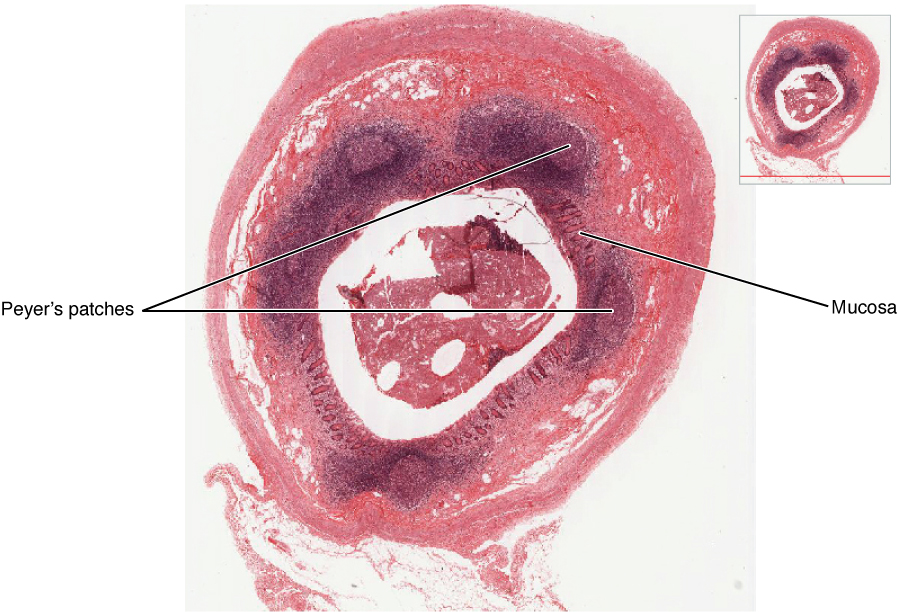
Find the rounded lymphoid tissue beneath the intestinal surface. Its position puts immune cells close to material arriving at the mucosa. This is a nodule within a tissue wall, not a separate encapsulated lymph node.
Image source and licence
OpenStax College · Source image · CC BY 3.0. Original image, reproduced without alteration.
Named nodules and where they sit
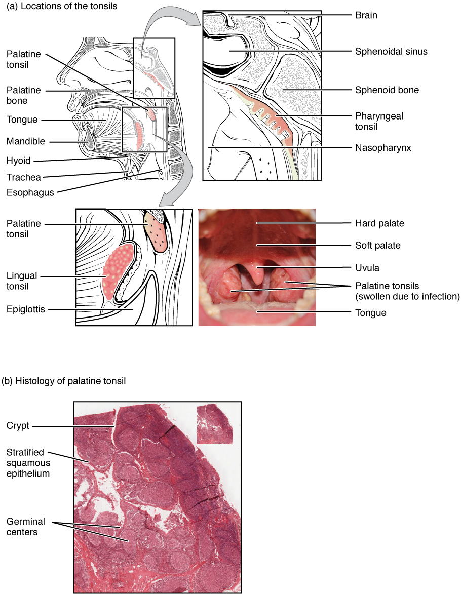
Several groups of nodules have their own names. The tonsils form a ring of lymphatic tissue around the entrance to the throat. The palatine tonsils sit on each side of the back of the mouth; the pharyngeal tonsil, or adenoid, lies in the roof of the nasopharynx; and the lingual tonsil sits at the base of the tongue. Together they guard the airway and food passages. In the small intestine, aggregated lymphoid nodules called Peyer's patches lie in the wall of the ileum, where they sample antigens from gut contents. The appendix contains a concentration of similar tissue. The airways have bronchus-associated lymphoid tissue, and the urinary tract has nodules in the bladder wall. Each group is positioned at a point where the body is most exposed to a particular environment.
For example: The palatine tonsils are visible at the back of the mouth; when they are inflamed the throat is sore and swallowing is painful.
 Enlarge illustration ↗
Locate the tonsils around the entrances to the digestive and respiratory passages. Then find the folds or crypts in the tissue view. Their surface position helps bring inhaled or swallowed material near immune tissue. The mouth photograph shows swollen, infected palatine tonsils, not their normal resting appearance.
Image source and licence
OpenStax College · Source image · CC BY 3.0. Original image, reproduced without alteration.
Why position and structure matter clinically
Because nodules sit at surfaces, they are often the first tissue to respond to an infection entering through that surface. Tonsillitis is inflammation of the tonsils, often with painful swallowing and enlarged tender tissue at the back of the throat. Inflammation of Peyer's patches can occur in gut infections, and the appendix, which contains similar tissue, can become inflamed and require removal. Because nodules have no capsule, inflammation spreads more freely through the surrounding tissue than it would from an encapsulated organ, and because they are not on lymphatic vessels, they do not produce the same pattern of lymph-borne spread as nodes. Recognising whether a finding is in a nodule or a node therefore helps explain its location and its clinical behaviour.
For example: Appendicitis causes pain that starts vaguely around the navel and localises to the right lower quadrant as the inflammation spreads through the wall of the appendix.
Key facts to remember
- A lymphatic nodule is an unencapsulated cluster of lymphocytes embedded in tissue, not a discrete organ.
- A lymph node is encapsulated and lies along a lymphatic vessel; a nodule has no capsule and sits within a tissue.
- Mucosa-associated lymphoid tissue (MALT) places immune tissue where microbes are most likely to enter.
- Named nodules include the palatine, pharyngeal and lingual tonsils, Peyer's patches of the ileum and the lymphoid tissue of the appendix.
- Nodules respond to locally arriving antigens rather than filtering flowing lymph, and their inflammation spreads through surrounding tissue.
Example · Compare two collections of lymphatic tissue. A cervical lymph node is a bean-shaped organ with a complete capsule, lying on a lymphatic vessel; it filters lymph arriving from the throat through afferent vessels and can be felt as a tender lump when it responds to infection. A palatine tonsil is a cluster of nodules without a capsule, sitting directly in the mucous membrane at the back of the mouth; it samples antigens from what is breathed and swallowed and becomes inflamed in tonsillitis. Both contain B and T cells and both enlarge when active, but their structure and position explain why one filters lymph and the other guards a surface.
Read full notes
Read one topic at a time. These notes explain the mechanisms and relationships behind the key facts.
Unencapsulated lymphatic tissue
The absence of a capsule has practical consequences. A lymph node can be dissected out as a discrete organ and its internal regions identified; a nodule merges with the tissue around it and is defined mainly by its cell population. Because a nodule has no afferent or efferent lymphatic vessels, lymph does not flow through it; instead, antigens reach it by diffusing through the surrounding tissue or by being carried there by immune cells. This makes nodules well suited to guarding surfaces, where antigens arrive directly, and poorly suited to filtering a stream of lymph, which is the node's role. Structure and function are matched in the same way as elsewhere in the body.
Nodules compared with nodes
It helps to compare the two along the same lines used for other tissues. Shape and boundary: a node is bean-shaped and sharply defined by its capsule; a nodule is rounded and blends into surrounding tissue. Internal organisation: a node has a cortex, paracortex and medulla; a nodule has a simpler arrangement of lymphocytes around a reticular network. Connection: a node is connected to afferent and efferent lymphatic vessels; a nodule is not. Position: nodes lie in regional drainage stations; nodules lie in the walls of organs that face the environment. Response: both enlarge when activated, but a node's enlargement is part of a lymph-borne response, while a nodule's is part of a local surface response.
MALT: defence at the surfaces
MALT is a strategy as much as a tissue. The body could rely on lymph nodes alone, but a microbe entering the gut would then have to travel in lymph before meeting organised immune tissue. By placing nodules directly in the gut wall, the airways and the urinary tract, the body intercepts antigens at the point of entry. MALT also contains specialised cells that sample antigens from the surface and deliver them to the nodules, and it produces antibodies, particularly of the IgA class, that are secreted back onto the surface. This arrangement is efficient: fewer microbes reach deeper tissues, and the response begins where it is needed. It explains why these surfaces have their own immune tissue rather than depending only on distant nodes.
Named nodules and where they sit
The named groups of nodules are positioned according to what they guard. The tonsils form a ring at the crossing point of the airway and the food passage, so they encounter both breathed and swallowed material. Peyer's patches lie in the ileum, the last part of the small intestine, where the contents are rich in bacteria and undigested material. The appendix, attached to the caecum, contains abundant lymphoid tissue near the beginning of the large intestine. Airway nodules sit in the bronchial walls, and bladder nodules in the urinary tract. In each case the location is not random: it is the point at which a particular surface is most exposed, and therefore where immune surveillance is most valuable.
Clinical relevance
Nodules produce familiar clinical problems. Tonsillitis causes a sore throat and painful swallowing, and in repeated severe cases the tonsils may be removed. Inflammation of Peyer's patches accompanies some gut infections, and the appendix, rich in similar tissue, can become inflamed and require surgery. Because nodules are unencapsulated and lie in the wall of an organ, inflammation can spread through the wall and even threaten it, which is why appendicitis is treated as an emergency. Recognising the difference between a nodule and a node also matters when interpreting imaging and biopsy reports, because the two structures have different drainage relationships and different patterns of involvement in disease.
Explore the diagram
Start with the whole view, then select a numbered structure. Compare the additional illustrations to connect what a structure looks like with what it does.
A lymphatic nodule beneath a mucosal surface
Enlarged generic mucosa, not a lymph node. A nodule is a local lymphocyte collection without a complete enclosing capsule.
A lining separates the lumen from internal tissue. Mucosal immune tissue lies close to places where material from outside meets the body.
Through a lymph node
Schematic cutaway. Several afferent vessels enter the convex surface; an efferent vessel leaves at the hilum. Trace the green flow arrows.
Lymph arrives from tissues through several vessels and enters the space beneath the capsule. Afferent means carrying toward the node.
Connect the illustrations to the key ideas
NODULE STRUCTURE · unencapsulated lymphocytes in tissue
A reticular network supports the cells; active nodules show a germinal centre.
MALT LOCATIONS · tonsils, Peyer's patches, appendix, airways, bladder
Nodules are placed where a surface meets the environment.
NODULE VERSUS NODE · no capsule, no afferent lymph; node is encapsulated
A nodule guards a surface; a node filters lymph flowing along a vessel.
Core Concepts Defined
- Lymphatic nodule (follicle)
- An unencapsulated cluster of lymphocytes embedded in tissue.
- Germinal centre
- The paler active centre of a nodule or follicle where B cells multiply.
- Reticular fibre
- A fine fibre forming the supporting network of lymphatic tissue.
- Mucosa-associated lymphoid tissue (MALT)
- Lymphatic tissue, including nodules, guarding mucous membranes.
- Tonsil
- A group of lymphatic nodules at the entrance to the throat.
- Palatine tonsil
- A tonsil on each side at the back of the mouth.
- Pharyngeal tonsil (adenoid)
- A tonsil in the roof of the nasopharynx.
- Lingual tonsil
- A tonsil at the base of the tongue.
- Peyer's patches
- Aggregated lymphoid nodules in the wall of the ileum.
- Appendix
- A small pouch of the large intestine containing abundant lymphoid tissue.
- Bronchus-associated lymphoid tissue
- Nodules guarding the walls of the airways.
- Tonsillitis
- Inflammation of the tonsils, often with sore throat and painful swallowing.
Go deeper · worked example
Nodule or node? Reading a lump
- Ask whether it is encapsulated. A lymph node has a complete fibrous capsule and a defined bean shape; a nodule has no complete capsule and merges with surrounding tissue.
- Ask whether it lies on a lymphatic vessel. Nodes are inserted into the lymphatic pathway and receive lymph through afferent vessels; nodules sit within a tissue and receive no afferent lymph.
- Consider the location. Nodules cluster where a surface meets the environment: the throat, the gut, the airways and the urinary tract. Nodes are found in regions such as the neck, armpit, groin and mediastinum, along drainage routes.
- Ask what it is responding to. A node responds to antigens carried from a distance in lymph; a nodule responds to antigens arriving locally from the surface it guards.
- Use the answer clinically. Encapsulated and on a vessel suggests a lymph node and a pattern of lymph-borne spread; unencapsulated and at a surface suggests a nodule, with inflammation spreading through the surrounding tissue instead.
Explain it yourself · Explain why a tonsil is not simply a lymph node in the throat.
Reveal a model explanation
A tonsil is a collection of lymphatic nodules, and a nodule differs from a node in three key ways. It has no complete fibrous capsule, so it is not a discrete encapsulated organ. It does not lie on a lymphatic vessel and receives no afferent lymph, so it does not filter flowing lymph. And it sits directly in the mucous membrane at the entrance to the throat, where it samples antigens from what is breathed and swallowed. A lymph node, by contrast, is a capsulated organ positioned along a lymphatic vessel, filtering lymph that arrives through afferent vessels. The tonsil guards a surface; the node filters a stream.
Check
Answer three questions from this section — one easy, one medium and one hard. If you miss one, you get a fresh set with explanations for the last attempt.
Practise this sectionOpen all questions and filter by difficulty or Bloom levelReferences and review status
OpenStax, Anatomy and Physiology 2e, 21.1 Anatomy of the Lymphatic and Immune Systems; Tortora & Derrickson, Principles of Anatomy and Physiology, 12e, T12-22A. Author review only; medical and French-language expert review pending.
Original teaching schematics. English and French wording is author-reviewed for study; anatomy and French-language expert review is pending. Checks are practice only, not a graded examination.
Practice progress is saved on this device for the current learner.