SECTION 7.3
Lymph nodes
Explain the structure of a lymph node, the direction lymph travels through it, and the filtering and immune work done there.
Learn
A lymph node is a small, bean-shaped organ placed along a lymphatic vessel, and it is one of the most important filters in the body. Each node has a fibrous capsule, an outer cortex containing clusters of immune cells called follicles, a deeper paracortex and a central medulla. Lymph enters through several afferent vessels on the convex surface, flows slowly through a network of sinuses where it is inspected and cleaned, and leaves through a single efferent vessel at the indented hilum. The slow flow gives macrophages time to remove particles and microbes, and it gives lymphocytes time to detect antigens and begin an immune response. This section explains the structure of a node, the one-way path of lymph through it, and why nodes enlarge and become tender in infection.
Key Concepts & Topics
A filter placed in the pathway
Lymph nodes are not scattered beside the lymphatic vessels; they are inserted into the pathway, so lymph from a region must pass through at least one node before returning to the blood. A node is usually only a few millimetres to about two centimetres long, and there are hundreds of them in the body, grouped in regions such as the neck, armpit, groin, mediastinum and abdomen. Because they sit in the drainage route, they perform two jobs at once: they filter the lymph, removing particles and microbes, and they provide a place where immune cells can meet antigens from the tissues. This double role explains why nodes are enlarged and tender in infection, and why cancer cells spreading through lymph often lodge in nodes first.
For example: The cervical nodes in the neck filter lymph from the throat, which is why a sore throat often causes tender lumps under the jaw.
Structure of a lymph node
A lymph node is covered by a fibrous capsule from which extensions called trabeculae pass inward, dividing the interior into compartments. Beneath the capsule is the cortex, the outer region, which contains rounded clusters of cells called follicles. Each follicle is rich in B lymphocytes; when the node is active, the centre of a follicle becomes a lighter germinal centre where B cells multiply and mature into antibody-producing cells. Deeper inside lies the paracortex, which is rich in T lymphocytes and in dendritic cells that present antigens. The central region is the medulla, containing macrophages and plasma cells in a network of sinuses. The indented side of the node is the hilum, where blood vessels enter and the efferent lymphatic vessel leaves.
For example: An active germinal centre is visible under the microscope as a paler centre in a follicle, showing that B cells are multiplying in response to an antigen.
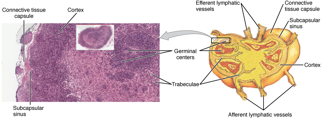 Enlarge illustration ↗
Match the tissue photograph to the organised cutaway. Look for the outer cortex and the deeper medulla, then locate the hilum. The drawing separates boundaries that are less obvious in real tissue.
Image source and licence
OpenStax College · Source image · CC BY 3.0. Original image, reproduced without alteration.
The path of lymph through a node
Lymph enters a node through several afferent vessels that pierce the convex surface. It then enters the subcapsular sinus, a space just beneath the capsule, and flows through the cortex and medulla along a network of sinuses. The sinuses are wide and contain many macrophages, so lymph slows as it passes and its particles are removed. Finally, the filtered lymph collects and leaves through a single efferent vessel at the hilum. This arrangement is one-way: afferent vessels enter on the convex surface and the efferent vessel leaves at the hilum, so lymph cannot bypass the filtering tissue. Because there are several afferent vessels but only one efferent vessel, flow slows further, which gives immune cells more time to encounter antigens.
For example: Because afferent vessels outnumber the efferent vessel, lymph slows in the node; this is a design feature, not an inefficiency.
Immune work in the node
The node is not only a filter but an immune command post. Dendritic cells that have picked up antigens in the tissues travel to the node and present those antigens to T lymphocytes in the paracortex. Activated T cells help B cells in the follicles, which multiply in germinal centres and produce antibodies. Some B cells become plasma cells that release antibodies into the lymph, and some become memory cells that persist for years. Macrophages in the sinuses engulf microbes and debris. This is why a node that is responding to an infection enlarges: immune cells multiply, blood flow increases and fluid accumulates, producing a swollen, often tender node. The tenderness reflects the rapid expansion and inflammation, and it usually subsides as the infection resolves.
For example: After vaccination, nodes near the injection site may enlarge for a few days because immune cells are multiplying in response to the vaccine antigen.
Enlarged nodes in practice
Enlarged lymph nodes, called lymphadenopathy, are a common clinical finding with several possible meanings. In an acute infection, nodes are usually tender, mobile and return to normal when the infection resolves. In cancer, nodes may be firm, non-tender and fixed to surrounding tissue, because tumour cells have replaced normal structure. Nodes may also enlarge in autoimmune conditions and in response to some drugs. The pattern matters: which nodes are enlarged suggests which region is draining into them. In cancer surgery, the first node to receive lymph from a tumour is called the sentinel node; identifying and examining it helps determine whether the cancer has begun to spread. This is why the anatomy of drainage routes and node groups is clinically important.
For example: A hard, fixed, painless node in the neck of a patient with a smoking history raises concern for cancer, whereas a tender mobile node usually suggests infection.
Key facts to remember
- Lymph nodes are inserted into the lymphatic pathway, so lymph from a region must pass through at least one node.
- A node has a fibrous capsule, a cortex with follicles rich in B cells, a paracortex rich in T cells, and a medulla with macrophages and plasma cells.
- Lymph enters through afferent vessels on the convex surface, flows through sinuses, and leaves through one efferent vessel at the hilum.
- Macrophages in the sinuses remove particles and microbes, while lymphocytes detect antigens and mount an immune response.
- Enlarged tender mobile nodes usually suggest infection; firm fixed non-tender nodes raise concern for cancer; the sentinel node is the first to receive lymph from a tumour.
Example · You develop a throat infection. Lymph from the throat passes through the cervical nodes, carrying bacteria and their antigens. Macrophages in the sinuses engulf microbes, while dendritic cells present antigens to T cells in the paracortex. T cells help B cells in the follicles, which multiply in germinal centres and produce antibodies. The node enlarges and becomes tender because immune cells are multiplying, blood flow is increasing and fluid is accumulating. Within days the infection resolves, cell numbers fall, and the node returns to its normal size.
Read full notes
Read one topic at a time. These notes explain the mechanisms and relationships behind the key facts.
A filter placed in the pathway
The strategic placement of lymph nodes is what makes them so useful. If they sat beside the vessels, lymph could bypass them; because they are inserted into the route, all lymph from a region must pass through. This means that particles, microbes and tumour cells travelling in lymph are exposed to the node's filtering and immune machinery before they can reach the general circulation. It also means that the node is an early warning system: changes there often precede or accompany disease in the tissue it drains. When a clinician examines a patient, the location of enlarged nodes is therefore a clue to the region that is affected.
Structure of a lymph node
The internal structure of a node reflects its two jobs. The cortex, with its follicles, is organised for B-cell responses and antibody production; the paracortex is organised for T-cell responses and antigen presentation; and the medulla, with its macrophages and plasma cells, is organised for filtration and antibody release. The capsule and trabeculae give the organ a framework and help direct lymph through the sinuses rather than letting it pass straight through. Blood vessels enter at the hilum to supply the metabolically active immune cells. Each region of the node therefore has a distinct role, and a biopsy can show which region is active, which helps distinguish different types of immune response.
The path of lymph through a node
The path of lymph is deliberately slow and convoluted. Entering through several afferent vessels, lymph first fills the subcapsular sinus and then percolates through the cortical and medullary sinuses before leaving by a single efferent vessel. Because the sinuses are wide and lined with macrophages, particles are removed as the fluid moves. Because there are many afferents and only one efferent, the flow slows, which increases the chance that an antigen will meet a lymphocyte. This is a good example of structure serving function: the anatomy of the node is designed to maximise contact between lymph and immune cells, and to give phagocytes time to work.
Immune work in the node
The immune work of a node is a coordinated sequence. Antigen-presenting cells arrive from the tissues and interact with T cells in the paracortex. Activated T cells migrate to the follicles and help B cells that have recognised the same antigen. B cells multiply in germinal centres and differentiate into antibody-secreting plasma cells and long-lived memory cells. Antibodies enter the lymph and eventually the blood, where they can act throughout the body. Memory cells remain so that a future exposure produces a faster, stronger response. This is why the node is described as a site of immune education, and why its enlargement is a sign that this education is actively taking place.
Enlarged nodes in practice
Because nodes are involved in so many processes, enlargement alone does not indicate a specific cause. The accompanying features help. Tender, mobile nodes that appear with a sore throat, a skin infection or a cold usually reflect a reactive immune response and settle on their own. Firm, fixed, non-tender nodes, especially when accompanied by weight loss or night sweats, raise concern for cancer and need prompt assessment. Nodes may also enlarge in autoimmune diseases, in response to some medicines and in rare conditions. In cancer care, the sentinel node concept uses the anatomy of drainage to find the first node a tumour is likely to reach, which allows a more targeted biopsy and can spare other nodes from removal.
Explore the diagram
Start with the whole view, then select a numbered structure. Compare the additional illustrations to connect what a structure looks like with what it does.
Through a lymph node
Schematic cutaway. Several afferent vessels enter the convex surface; an efferent vessel leaves at the hilum. Trace the green flow arrows.
Lymph arrives from tissues through several vessels and enters the space beneath the capsule. Afferent means carrying toward the node.
Connect the illustrations to the key ideas
NODE STRUCTURE · capsule, cortex with follicles, paracortex, medulla
B cells cluster in follicles; T cells and dendritic cells occupy the paracortex.
FLOW PATH · afferent vessels → sinuses → one efferent vessel
Lymph slows and is filtered; flow is one-way from convex surface to hilum.
FILTER AND IMMUNE ROLE · macrophages remove; lymphocytes respond
Particles and microbes are removed while antigens trigger antibody and memory responses.
Core Concepts Defined
- Lymph node
- A small bean-shaped organ along a lymphatic vessel that filters lymph and supports immune responses.
- Capsule
- The fibrous outer covering of a lymph node.
- Trabecula
- An inward extension of the capsule that divides the node into compartments.
- Cortex
- The outer region of a lymph node, containing follicles rich in B cells.
- Follicle
- A rounded cluster of lymphocytes, mainly B cells, in the node cortex.
- Germinal centre
- The paler active centre of a follicle where B cells multiply and mature.
- Paracortex
- The region deep to the cortex, rich in T cells and dendritic cells.
- Medulla
- The central region of a lymph node, containing macrophages and plasma cells in sinuses.
- Sinus
- A channel within a lymph node through which lymph flows slowly and is filtered.
- Subcapsular sinus
- The sinus just beneath the capsule, the first space lymph enters.
- Hilum
- The indented side of a node where the efferent vessel and blood vessels enter or leave.
- Afferent vessel
- A vessel bringing lymph into a node through its convex surface.
- Efferent vessel
- The single vessel carrying filtered lymph out of a node at the hilum.
- Macrophage
- A phagocytic cell that engulfs particles and microbes, abundant in node sinuses.
- Dendritic cell
- A cell that captures antigens and presents them to T lymphocytes in the node.
- Lymphadenopathy
- Enlargement of lymph nodes, from infection, cancer or other causes.
- Sentinel node
- The first lymph node to receive lymph from a tumour.
Go deeper · worked example
Why neck nodes swell with a sore throat
- Lymph carries the problem to the node. Infection in the throat releases bacteria and their antigens into the tissue fluid, which becomes lymph and travels to the cervical nodes.
- Macrophages filter. Lymph enters through afferent vessels, slows in the sinuses, and macrophages engulf microbes and debris, so fewer particles continue onward.
- Dendritic cells present antigen. Dendritic cells that have captured antigens travel to the paracortex and present them to T lymphocytes.
- B cells respond. Activated T cells help B cells in the follicles, which multiply in germinal centres and produce antibodies specific to the microbe.
- The node enlarges and then settles. The multiplication of immune cells, increased blood flow and fluid accumulation make the node swollen and tender; as the infection resolves, cell numbers fall and the node returns to normal size.
Explain it yourself · Explain why a lymph node enlarges during an infection and why it usually returns to normal afterwards.
Reveal a model explanation
When lymph carrying microbes and antigens arrives, the node mounts an immune response. Dendritic cells present antigen to T cells, T cells help B cells, and B cells multiply in germinal centres. Macrophages become more active and blood flow to the node increases. The combination of more immune cells, more blood and more fluid makes the node swell, and inflammation makes it tender. Once the infection is controlled, the stimulus for cell multiplication disappears, cell numbers fall and fluid is reabsorbed, so the node returns to its usual size. A node that stays enlarged, hard and non-tender does not fit this pattern and needs further assessment.
Check
Answer three questions from this section — one easy, one medium and one hard. If you miss one, you get a fresh set with explanations for the last attempt.
Practise this sectionOpen all questions and filter by difficulty or Bloom levelReferences and review status
OpenStax, Anatomy and Physiology 2e, 21.1 Anatomy of the Lymphatic and Immune Systems; Tortora & Derrickson, Principles of Anatomy and Physiology, 12e, T12-22A. Author review only; medical and French-language expert review pending.
Original teaching schematics. English and French wording is author-reviewed for study; anatomy and French-language expert review is pending. Checks are practice only, not a graded examination.
Practice progress is saved on this device for the current learner.