SECTION 7.1
Formation and entry of lymph
Explain how interstitial fluid becomes lymph and how it enters a lymphatic capillary without flowing straight back out.
Learn
Every day, fluid leaves the blood capillaries to bathe the cells. Most of it returns to the capillaries, but some does not, and that leftover fluid would gradually accumulate and cause swelling if nothing removed it. The lymphatic system solves this problem. The fluid in the tissue spaces is called interstitial fluid; once it enters a lymphatic vessel, the same fluid is called lymph. Lymphatic capillaries are blind-ended tubes that begin in the tissue spaces, and their walls are built so that fluid can enter easily but cannot flow straight back out. This section explains how fluid leaves the blood, how it becomes lymph, and how a simple overlapping-cell mechanism makes entry one-way. It is the first step in understanding fluid balance, immunity and the swelling that appears when drainage fails.
Key Concepts & Topics
Why fluid leaves the blood capillaries
At the arterial end of a capillary, blood pressure pushes water and small dissolved substances out through the vessel wall into the tissue space. This movement is called filtration, and it delivers oxygen, nutrients and water to the cells. At the venous end, the pressure inside the capillary is lower and the osmotic pull of plasma proteins draws most of the fluid back in. In a healthy person, the two movements nearly balance: most of the filtered fluid is reabsorbed. A small fraction, perhaps two to four litres a day, is not, and it remains in the tissue spaces. If it were allowed to accumulate, the tissues would swell. The lymphatic system exists to collect this residual fluid and return it to the blood.
For example: When you stand for a long time, capillary pressure in the legs is higher, more fluid filters out and the ankles may swell slightly until lymph drainage catches up.
Interstitial fluid becomes lymph
The fluid in the tissue spaces is interstitial fluid. It contains water, dissolved ions and nutrients, small proteins that have leaked from the capillaries, waste products from cell activity and sometimes particles such as bacteria. When this fluid enters a lymphatic capillary, it is called lymph. The change of name marks the change of compartment, not a change in the fluid itself: interstitial fluid is outside the vessels, and lymph is inside a lymphatic vessel. Lymph is normally clear or pale yellow and similar in composition to plasma but with much less protein. Because it comes from the tissue spaces, its composition reflects what is happening locally; lymph from the intestine is rich in absorbed fats, and lymph from an infected area contains microbes and immune cells.
For example: Lymph collected from the small intestine after a fatty meal looks milky, because it carries absorbed fat; lymph elsewhere is clear.
How lymph enters a lymphatic capillary
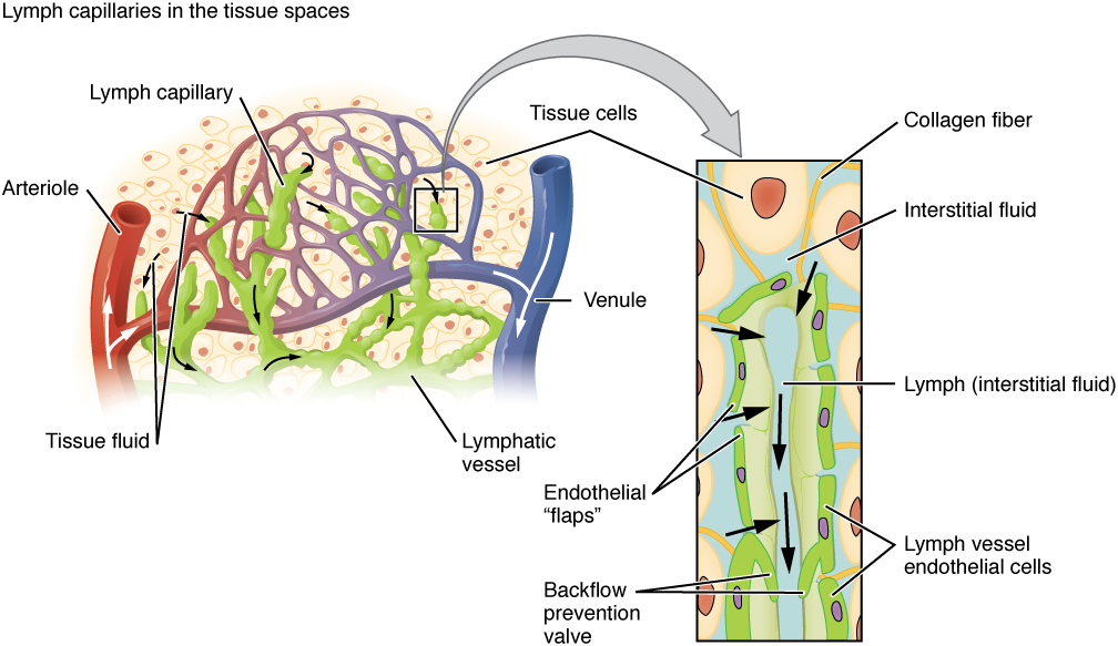
Lymphatic capillaries are blind-ended: one end is closed, and the open end continues into larger vessels. They begin in the tissue spaces, so their closed tips lie close to the cells. Their walls are made of endothelial cells that overlap one another rather than forming a sealed tube, like tiles on a roof. When pressure in the tissue fluid rises above the pressure inside the capillary, the overlapping edges are pushed apart and fluid enters through the gaps. Fine anchoring filaments connect the endothelial cells to the surrounding tissue; when the tissue swells, the filaments pull the cells apart and widen the openings, so more fluid can enter exactly when it is most needed. This is a passive, pressure-driven mechanism that requires no pump.
For example: If excess fluid makes a tissue swell, the anchoring filaments are stretched and pull the capillary cells apart, so the entry openings widen automatically.
 Enlarge illustration ↗
Follow fluid from the tissue space into the blind-ended lymphatic capillary. The overlapping cell edges act as entry flaps. Once interstitial fluid has entered this vessel, it is called lymph; it has not turned into a different substance.
Image source and licence
OpenStax College · Source image · CC BY 3.0. Original image, reproduced without alteration.
Why lymph does not flow straight back out
The same overlapping-cell design that lets fluid in also prevents it from leaving. Once fluid has entered, pressure inside the capillary rises. That internal pressure pushes the overlapping edges of the endothelial cells together, closing the gaps from the inside, rather than apart. The result is a one-way valve made of the wall itself: fluid can enter when outside pressure is higher, and backflow is blocked when inside pressure is higher. Larger lymphatic vessels add conventional valves along their length, which keep lymph moving in one direction as it is squeezed by body movements. This combination means that lymph moves from the tissue spaces towards the centre of the body and does not return to the spaces it came from.
For example: If you press on a swollen area, you do not push lymph back into the tissue; the overlapping cell edges close against the pressure.
Common misconception: Lymphatic capillaries are not open pipes. Their walls are sealed by overlapping cells that open only when outside pressure exceeds inside pressure.
Where lymphatic capillaries are found
Lymphatic capillaries are present in most tissues, but not all. They are absent from the epidermis, from cartilage and the cornea, and from the central nervous system, which has its own fluid drainage arrangements. Where they are present, they lie close to blood capillaries so that they can collect the fluid that filters out. In the small intestine, specialised lymphatic capillaries called lacteals extend into each villus and absorb fats and fat-soluble vitamins that cannot enter the blood capillaries directly. Because lymphatic capillaries depend on tissue pressure and movement rather than on the heart, drainage is slow and low-pressure, and it depends on the skeletal muscle pump, breathing movements and the pulsation of nearby arteries to keep lymph moving.
For example: A scratch on the cornea heals without lymphatic drainage because the cornea has no lymphatic capillaries; the aqueous humour and other routes handle fluid.
Key facts to remember
- Fluid leaves blood capillaries by filtration; most is reabsorbed, and the remainder becomes interstitial fluid.
- Interstitial fluid becomes lymph when it enters a lymphatic capillary; the fluid is the same, only the compartment changes.
- Lymphatic capillaries are blind-ended tubes with overlapping endothelial cells and anchoring filaments.
- Rising outside pressure opens the overlaps and lets fluid in; rising inside pressure closes them and prevents backflow.
- Lymphatic capillaries are absent from epidermis, cartilage, cornea and the central nervous system; lacteals in the intestine absorb fats.
Example · You sprain your ankle. The injury makes capillaries leak more fluid than usual, so interstitial fluid accumulates and the ankle swells. As the tissue pressure rises, the anchoring filaments pull the lymphatic capillary cells apart and lymph enters more readily. The fluid is carried away towards larger vessels and eventually returned to the blood. If the injury is severe or you keep the ankle dependent, the fluid forms faster than it can be removed and the swelling increases. Elevating the ankle lowers capillary pressure, so less fluid filters out and the existing lymph can drain.
Read full notes
Read one topic at a time. These notes explain the mechanisms and relationships behind the key facts.
Fluid in the tissue space
The tissue space is not empty: it contains a film of fluid that bathes every cell. This fluid is the cell’s immediate environment, and its composition determines what reaches the cell membrane. It is produced by filtration from blood capillaries and modified by the cells themselves, which take up nutrients and release waste. Because it is constantly renewed, it is also constantly removed: some returns to the blood capillaries by reabsorption, and the rest enters lymphatic capillaries. The balance between filtration, reabsorption and lymphatic drainage determines how much fluid remains. If filtration increases, if reabsorption falls, or if lymphatic drainage is blocked, fluid accumulates and oedema appears. This is why the first step in understanding swelling is to ask which of the three has changed.
Interstitial fluid becomes lymph
The change from interstitial fluid to lymph is a change of compartment, not of substance. The fluid is the same water-based solution; what changes is that it has crossed the wall of a lymphatic capillary and is now inside the lymphatic system. This matters because the two compartments behave differently. Interstitial fluid exerts pressure on the cells around it and contributes to tissue swelling; lymph is being transported away and no longer contributes to tissue pressure. Naming the fluid by its compartment is therefore not a technicality but a way of tracking where it is and what it is doing. Once lymph has entered the system, it travels through vessels and nodes towards the trunk and is eventually returned to the venous blood.
The one-way entry mechanism
The one-way mechanism is an elegant example of structure producing function without energy cost. Because the endothelial cells overlap rather than seal, the wall is a kind of valve that responds to whichever side has more pressure. This is why lymphatic capillaries collect fluid even though they have no pump of their own: they rely on the pressure of the tissue fluid around them. It also explains why lymphatic drainage improves when the tissue is swollen and worsens when the tissue is compressed from outside, such as by tight clothing or prolonged pressure. The anchoring filaments add a useful refinement: they couple the opening of the gaps to the swelling of the tissue, so the system responds automatically to the very condition it exists to relieve.
Where lymphatic capillaries are found
The distribution of lymphatic capillaries follows their function. They are present where fluid filters out of blood capillaries and where particles may enter from the environment or from the gut. They are absent from tissues with no blood capillaries of the usual kind, such as the epidermis and cartilage, and from the central nervous system, which maintains its own fluid environment. Their absence has consequences: because the cornea and cartilage have no lymphatic drainage, fluid and debris are handled by other routes, and an injury there heals differently. Their presence in the gut, as lacteals, explains why fats and fat-soluble vitamins are absorbed into lymph rather than directly into the blood, which is why lymph from the intestine appears milky after a fatty meal.
What happens when entry fails
If lymphatic capillaries fail to collect interstitial fluid, the fluid accumulates and the tissue swells. This can happen for several reasons. The lymphatic vessels may be blocked, as by a tumour or by surgical removal of nodes; the valves or vessel walls may be damaged; or the volume of filtered fluid may simply exceed what the lymphatic system can remove, as in heart or kidney failure. Because lymph also carries immune cells and particles, blocked drainage can impair local defence as well as cause swelling. Understanding the entry step is therefore the basis for interpreting swelling and for treatments such as compression, elevation, movement and massage, which all aim to help fluid move from the tissue space into the lymphatic system.
Explore the diagram
Start with the whole view, then select a numbered structure. Compare the additional illustrations to connect what a structure looks like with what it does.
Lymph drainage and the lymphatic organs
Anterior schematic view, not to scale. Select a number below to trace one connection at a time. The patient’s right is on your left. Green marks lymphatic routes; purple marks lymphoid tissue.
More controls
01 / 18 Initial lymphatic capillaries
A closed lymphatic capillary
Initial lymphatic capillaries
These blind-ended vessels begin in most tissues. Overlapping cells in their wall let interstitial fluid enter, and anchoring filaments open the gaps when the tissue swells. Fluid inside them is now called lymph.
Illustration credits
- Goran tek-en · Male front 3d-shaded human illustration.svg · CC BY-SA 4.0.

Adapted for this lesson: labels removed, artwork cropped or recoloured, and interactive teaching overlays added. Adapted artwork retains its source licence.
Connect the illustrations to the key ideas
FILTRATION OUT · blood capillary pressure pushes fluid out
Water, ions and small proteins enter the tissue space; most returns by reabsorption.
INTERSTITIAL FLUID · the fluid bathing the cells
What remains after reabsorption is the fluid the lymphatic system must collect.
ENTRY INTO LYMPH · overlapping cells open outward, close inward
Higher outside pressure opens the gaps; higher inside pressure seals them, so entry is one-way.
Core Concepts Defined
- Filtration
- Pressure-driven movement of water and small solutes out of a capillary into the tissue space.
- Reabsorption
- Return of fluid from the tissue space into the blood capillary, driven mainly by osmotic pull.
- Interstitial fluid
- The fluid in the spaces between cells, outside the blood and lymphatic vessels.
- Lymph
- Interstitial fluid once it has entered a lymphatic vessel.
- Lymphatic capillary
- A blind-ended vessel beginning in the tissue spaces that collects interstitial fluid.
- Blind-ended
- Closed at one end, as the initial lymphatic capillaries are.
- Overlapping endothelial cells
- The tile-like arrangement that lets fluid enter but blocks backflow.
- Anchoring filaments
- Fine fibres linking lymphatic endothelial cells to surrounding tissue so swelling widens the entry gaps.
- Lacteal
- A specialised lymphatic capillary in an intestinal villus that absorbs fats.
- Oedema
- Swelling caused by excess fluid in the tissue spaces.
- Skeletal muscle pump
- The action of moving muscles squeezing lymphatic and venous vessels to help fluid return.
Go deeper · worked example
Why an ankle swells after a sprain
- Fluid filters out. Injury makes the capillary wall more permeable and raises local capillary pressure, so more water and small proteins leave the blood than usual.
- The tissue space fills. The extra fluid raises the volume of interstitial fluid, and the tissue begins to swell.
- Anchoring filaments respond. As the tissue swells, the filaments linking lymphatic endothelial cells to the tissue are stretched, pulling the overlapping cell edges apart and widening the entry gaps.
- Lymph enters and is held. Because outside pressure is now higher than inside pressure, fluid enters the lymphatic capillary; once inside, the higher internal pressure presses the overlaps closed and prevents return.
- Drainage and treatment. Lymph is carried to larger vessels and eventually returned to the blood. Elevation lowers capillary pressure so less fluid filters out, and movement helps the muscle pump move the lymph, which is why both are used in treatment.
Explain it yourself · Explain why lymph can enter a lymphatic capillary but cannot immediately flow back out into the tissue.
Reveal a model explanation
The capillary wall is made of endothelial cells that overlap like roof tiles rather than forming a sealed tube. When tissue fluid pressure rises above the pressure inside the capillary, the overlapping edges are pushed apart and fluid enters. Once fluid is inside, the internal pressure rises above the tissue pressure, and that higher internal pressure presses the overlapping edges together, closing the gaps from within. The wall therefore acts as a one-way valve: outside pressure opens it, inside pressure shuts it. Larger lymphatic vessels add conventional valves, so lymph continues towards the centre of the body instead of returning to the tissue space.
Check
Answer three questions from this section — one easy, one medium and one hard. If you miss one, you get a fresh set with explanations for the last attempt.
Practise this sectionOpen all questions and filter by difficulty or Bloom levelReferences and review status
OpenStax, Anatomy and Physiology 2e, 21.1 Anatomy of the Lymphatic and Immune Systems; Tortora & Derrickson, Principles of Anatomy and Physiology, 12e, T12-22A. Author review only; medical and French-language expert review pending.
Original teaching schematics. English and French wording is author-reviewed for study; anatomy and French-language expert review is pending. Checks are practice only, not a graded examination.
Practice progress is saved on this device for the current learner.