SECTION 6.1
Heart location, membranes and chambers
Locate the heart in the thorax, name its membranes and chambers, and relate each chamber to the circuit it serves.
Learn
The heart is a muscular pump about the size of a closed fist, sitting in the centre of the chest behind the breastbone and between the lungs. It is tilted so that its pointed lower end, the apex, lies to the left, which is why people place a hand on the left side of the chest to feel it. The heart is wrapped in a protective sac called the pericardium and is divided internally into four chambers. The two upper chambers, the atria, receive blood; the two lower chambers, the ventricles, pump it out. The right side of the heart serves the pulmonary circuit and the left side serves the systemic circuit. This section locates the heart, describes the layers that surround and compose it, and introduces the four chambers and the great vessels connected to them.
Key Concepts & Topics
Where the heart sits
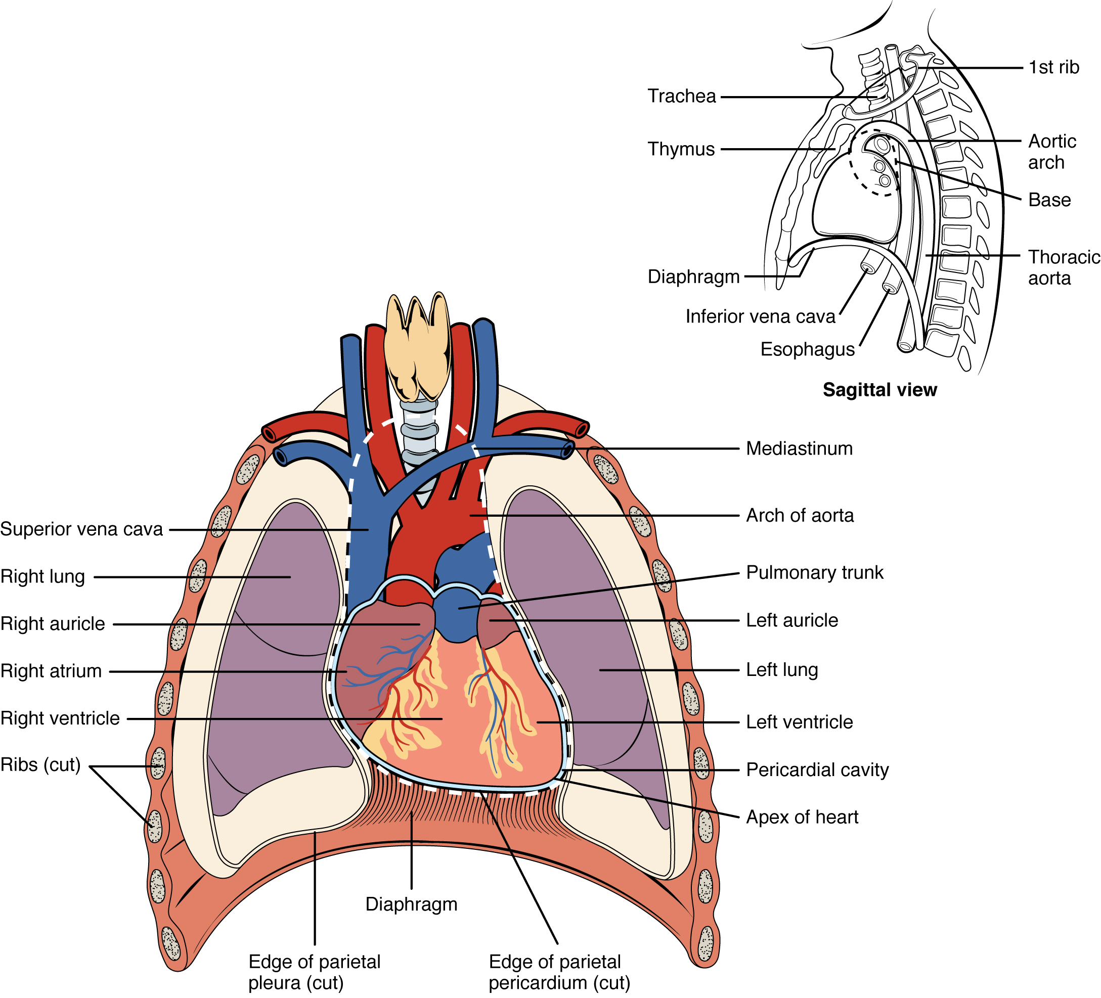
The heart lies in the mediastinum, the central compartment of the thoracic cavity between the two lungs. It sits behind the sternum and in front of the vertebral column, above the diaphragm, and roughly two-thirds of it lies to the left of the midline. Its broad upper part, the base, faces upward and to the right and is where the great vessels enter and leave; its pointed lower end, the apex, points down, forward and to the left. This orientation explains several clinical facts: the heartbeat is felt best at the apex, between the fifth and sixth ribs, and the heart sits close to the front of the chest, which is why it can be examined with a stethoscope, ultrasound and X-ray. Because it lies between the lungs, its position changes with breathing and posture.
For example: Placing a stethoscope at the apex, on the left side below the nipple line, gives the clearest sound of the mitral valve closing.
 Enlarge illustration ↗
Use the lungs and diaphragm as landmarks. The heart occupies the mediastinum between the lungs; it is not entirely in the left side of the chest. Its apex points toward the patient’s left.
Image source and licence
OpenStax College · Source image · CC BY 3.0. Original image, reproduced without alteration.
Pericardium and the heart wall
The heart is enclosed by the pericardium, a sac with two parts. The outer fibrous pericardium is tough connective tissue that anchors the heart to surrounding structures and prevents it from overfilling. Inside it, the serous pericardium forms two layers: the parietal layer lines the fibrous sac, and the visceral layer, also called the epicardium, is attached directly to the heart muscle. Between the two layers is the pericardial cavity, a thin space containing a small amount of serous fluid that lets the heart move without friction. The heart wall itself has three layers. The epicardium is the outer covering; the myocardium is the thick middle layer of cardiac muscle that does the pumping; and the endocardium is the smooth inner lining that contacts the blood and continues into the vessels. The myocardium is thickest in the left ventricle because that chamber pumps against the highest pressure.
For example: In pericarditis, the inflamed layers rub together and cause sharp chest pain, because the fluid that normally lets them glide is insufficient.
The four chambers
The heart has four chambers arranged in two pairs. The right atrium receives deoxygenated blood from the body through the superior vena cava, the inferior vena cava and the coronary sinus, which drains the heart’s own muscle. The right ventricle receives blood from the right atrium and pumps it into the pulmonary trunk towards the lungs. The left atrium receives oxygenated blood from the lungs through the pulmonary veins. The left ventricle receives blood from the left atrium and pumps it into the aorta towards the body. A septum separates the two sides: the interatrial septum between the atria and the interventricular septum between the ventricles. Small ear-like extensions of the atria, called auricles, increase their volume slightly. Because the left ventricle must generate systemic pressure, its muscular wall is much thicker than that of the right ventricle.
For example: A blood sample taken from the right atrium is deoxygenated, while one taken from the left atrium is oxygenated — the same heart, two different circuits.
Common misconception: The atria are the receiving chambers and the ventricles are the pumping chambers. Blood does not flow directly from the body into a ventricle.
The great vessels
Each chamber connects to specific great vessels, and knowing them makes blood flow easy to follow. Into the right atrium: the superior vena cava from the head, neck and arms; the inferior vena cava from the trunk and legs; and the coronary sinus from the heart muscle itself. Out of the right ventricle: the pulmonary trunk, which divides into the right and left pulmonary arteries. Into the left atrium: four pulmonary veins, two from each lung. Out of the left ventricle: the aorta, the largest artery, which arches and descends to supply the whole body. The pattern is simple once the circuits are understood: veins bring blood to the atria, and arteries carry it away from the ventricles. The only apparent exception, the pulmonary trunk carrying deoxygenated blood, follows the direction rule explained in section 5.2.
For example: The superior vena cava enters the right atrium from above and the inferior vena cava from below, which is why both are named for their position relative to the heart.
Chambers and the circuits they serve
The four chambers are best remembered by the circuit each serves. The right atrium and right ventricle form the right pump, which sends blood to the lungs; the left atrium and left ventricle form the left pump, which sends blood to the body. Because the two circuits are in series, the two pumps must move the same volume over time, which is why the right and left ventricles have matched outputs. Their walls differ because their workloads differ: the right ventricle pumps into the low-pressure pulmonary circuit, while the left pumps into the high-pressure systemic circuit. This is why the left ventricular wall is roughly three times thicker and why diseases that raise pressure in one circuit change the shape and workload of the corresponding ventricle.
For example: In pulmonary hypertension, the right ventricle must work against higher resistance and its wall thickens over time, just as the left ventricle does in systemic hypertension.
Key facts to remember
- The heart lies in the mediastinum, behind the sternum and between the lungs, with its apex pointing down and to the left.
- The pericardium has a fibrous outer part and a serous inner part with parietal and visceral layers separated by lubricating fluid.
- The heart wall has three layers: epicardium, myocardium and endocardium; the myocardium is thickest in the left ventricle.
- The right atrium and ventricle serve the pulmonary circuit; the left atrium and ventricle serve the systemic circuit.
- Veins bring blood to the atria (venae cavae, coronary sinus, pulmonary veins); arteries carry it from the ventricles (pulmonary trunk, aorta).
Example · Imagine following a drop of blood through the heart. It arrives from the legs in the inferior vena cava and enters the right atrium. It passes into the right ventricle and is pumped into the pulmonary trunk, then to a lung. After picking up oxygen, it returns in a pulmonary vein to the left atrium. From there it enters the left ventricle and is pumped into the aorta, which carries it to the body. Every chamber it visited had one job: the atria received, the ventricles pumped, the right side served the lungs and the left side served the body.
Read full notes
Read one topic at a time. These notes explain the mechanisms and relationships behind the key facts.
Where the heart sits
The heart’s position explains much of how it is examined and how it is affected by disease. It lies in the mediastinum, angled so that the apex points down and to the left. Because it is close to the front of the chest, its sounds are transmitted to the chest wall and can be heard with a stethoscope; the valves are heard best at specific points on the chest rather than directly over the heart. Because it lies between the lungs and above the diaphragm, its position shifts with breathing and with changes in posture. Because it is enclosed by the pericardium, it is protected from friction and from sudden overfilling, but that same enclosure means that fluid accumulating in the pericardial cavity can compress the heart and impair filling.
Membranes and wall layers
The membranes around the heart have a mechanical and a lubricating role. The fibrous pericardium is strong and largely inelastic; it holds the heart in place and resists sudden stretching. The serous pericardium provides a smooth, fluid-lubricated interface so that the beating heart does not rub against its surroundings. The heart wall layers each serve the pumping function. The epicardium is a thin outer covering that also contains some fat and vessels. The myocardium is the working muscle; its thickness varies with the pressure each chamber must generate. The endocardium is smooth and continuous with the lining of the vessels, which reduces friction and helps prevent clot formation inside the heart. Each layer therefore contributes to safe, efficient pumping rather than merely wrapping the organ.
The four chambers
The four-chamber design separates oxygenated from deoxygenated blood and lets each side serve one circuit. The atria are thin-walled because they only have to move blood a short distance into the ventricles; they act as reservoirs and as priming pumps. The ventricles are thick-walled because they must generate the pressure to push blood through a circuit. The right ventricle pumps into the low-resistance lungs, so its wall is thinner; the left ventricle pumps into the high-resistance body, so its wall is much thicker. The septum prevents mixing between the two sides, which is essential for efficient oxygen delivery. When the septum has an abnormal opening, oxygenated and deoxygenated blood mix, and the heart must work harder to compensate.
The great vessels
The great vessels are the connections between the heart and the circuits, and their arrangement follows a simple logic. Veins return blood to the atria from the body (venae cavae), the heart itself (coronary sinus) and the lungs (pulmonary veins). Arteries carry blood away from the ventricles to the lungs (pulmonary trunk) and to the body (aorta). Because the great vessels are named for direction, the pulmonary trunk is an artery even though it carries deoxygenated blood. Recognising this logic makes it possible to reconstruct the flow through the heart from a list of vessel names, and to predict what happens if a connection is abnormal, as in transposition of the great arteries.
Chambers and the circuits they serve
The heart is best understood as two pumps working in series, not one pump with four parts. The right pump receives from the body and sends to the lungs; the left pump receives from the lungs and sends to the body. Because the circuits are in series, each pump must move the same volume, so the two ventricles have matched outputs. Their different wall thicknesses reflect different pressures, not different volumes. Diseases usually affect one pump first: left-sided failure congests the lungs, while right-sided failure congests the systemic veins and produces swelling in the legs. This is why a clinician examining a patient with breathlessness or ankle swelling thinks in terms of which pump is failing, and why the anatomy of the chambers and their connections is the starting point for that reasoning.
Explore the diagram
Start with the whole view, then select a numbered structure. Compare the additional illustrations to connect what a structure looks like with what it does.
Anterior relationships
Reduced motion · use the slider to explore each state.
Anterior to the heart; the lungs lie on either side.
Educational sequence · slowed and simplified, not real-time physiology · Guided landmark tour
Inside the heart
Reduced motion · use the slider to explore each state.
Receives the venae cavae and coronary sinus.
Educational sequence · slowed and simplified, not real-time physiology
From blood to the pericardial sac
Reduced motion · use the slider to explore each state.
Smooth inner lining in contact with chamber blood.
Educational sequence · slowed and simplified, not real-time physiology · Layers separate for study; this is not tissue movement.
Connect the illustrations to the key ideas
LOCATION AND PERICARDIUM · mediastinum; sac with lubricating fluid
The apex points down and left; the pericardium anchors the heart and reduces friction.
FOUR CHAMBERS · two atria receive, two ventricles pump
The septum separates right from left; the left ventricular wall is thicker.
GREAT VESSELS · venae cavae and pulmonary veins in; trunk and aorta out
Veins enter the atria, arteries leave the ventricles; direction, not oxygen, names them.
Core Concepts Defined
- Mediastinum
- The central compartment of the thorax between the lungs, containing the heart and great vessels.
- Base of the heart
- The broad upper part of the heart where the great vessels enter and leave.
- Apex of the heart
- The pointed lower end of the heart, directed down, forward and to the left.
- Pericardium
- The sac surrounding the heart, with fibrous and serous parts.
- Fibrous pericardium
- The tough outer layer that anchors the heart and limits overfilling.
- Serous pericardium
- The inner double layer with parietal and visceral parts and lubricating fluid between them.
- Parietal layer
- The serous layer lining the fibrous pericardium.
- Visceral layer (epicardium)
- The serous layer attached directly to the heart muscle.
- Pericardial cavity
- The thin space between the serous layers, containing lubricating fluid.
- Myocardium
- The thick middle layer of cardiac muscle that performs the pumping.
- Endocardium
- The smooth inner lining of the heart that contacts the blood.
- Atrium
- An upper receiving chamber of the heart.
- Ventricle
- A lower pumping chamber of the heart.
- Interatrial septum
- The wall separating the two atria.
- Interventricular septum
- The wall separating the two ventricles.
- Auricle
- A small ear-like extension of an atrium that increases its volume.
- Superior vena cava
- The vein returning blood from the head, neck and arms to the right atrium.
- Inferior vena cava
- The vein returning blood from the trunk and legs to the right atrium.
- Coronary sinus
- The vessel that drains blood from the heart muscle into the right atrium.
- Pulmonary trunk
- The vessel leaving the right ventricle, dividing into the pulmonary arteries.
- Pulmonary veins
- The four veins returning oxygenated blood from the lungs to the left atrium.
- Aorta
- The largest artery, leaving the left ventricle to supply the body.
Go deeper · worked example
Which chamber is it?
- Decide which circuit the blood belongs to. Blood returning from the body is deoxygenated and belongs to the right side; blood returning from the lungs is oxygenated and belongs to the left side.
- Identify the receiving chamber. Blood always enters an atrium first. From the body it enters the right atrium through the venae cavae or the coronary sinus; from the lungs it enters the left atrium through the pulmonary veins.
- Identify the pumping chamber. Blood passes from the atrium to the ventricle on the same side: right atrium to right ventricle, left atrium to left ventricle.
- Identify the outlet vessel. The right ventricle ejects into the pulmonary trunk; the left ventricle ejects into the aorta. Direction, not oxygen content, tells you whether the vessel is an artery.
- Check against the rule. Atria receive, ventricles pump, right side serves the lungs and left side serves the body. Any answer that breaks this pattern should be re-examined.
Explain it yourself · A blood sample from a chamber is found to be oxygenated. Which chambers and vessels could it have come from, and why?
Reveal a model explanation
Oxygenated blood has passed through the lungs, so it belongs to the left side of the heart before it is used by the tissues. It could come from a pulmonary vein, the left atrium or the left ventricle. It could also be in the aorta or its branches once it has left the left ventricle. It cannot come from the venae cavae, the right atrium or the right ventricle, because those carry blood that has already delivered oxygen to the body. Knowing the circuits turns a single observation into a set of possible locations.
Check
Answer three questions from this section — one easy, one medium and one hard. If you miss one, you get a fresh set with explanations for the last attempt.
Practise this sectionOpen all questions and filter by difficulty or Bloom levelReferences and review status
OpenStax, Anatomy and Physiology 2e, 19.1 Heart Anatomy; Tortora & Derrickson, Principles of Anatomy and Physiology, 12e, ch. 20. Author review only; medical and French-language expert review pending.
Original teaching schematics. English and French wording is author-reviewed for study; anatomy and French-language expert review is pending. Checks are practice only, not a graded examination.
Practice progress is saved on this device for the current learner.