SECTION 9.3
Functions of the liver
Identify the main functions of the liver in metabolism, storage, detoxification and bile production.
Learn
The liver is the largest internal organ and one of the busiest. It sits in the upper right abdomen beneath the diaphragm and receives blood from two sources: the hepatic artery brings oxygenated blood, and the hepatic portal vein brings nutrient-rich blood from the intestines. Because everything absorbed from the gut passes through the liver before reaching the rest of the body, the liver can adjust nutrient levels, store what is not needed now and remove substances that must not circulate. Its main functions fall into four groups: metabolism of carbohydrates, fats and proteins; storage of glycogen, vitamins, iron and other materials; detoxification of drugs, alcohol and waste products; and production of bile, which is needed for fat digestion. This section explains these functions and what happens when the liver fails.
Key Concepts & Topics
Structure and blood supply
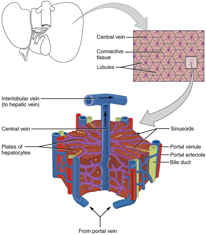
The liver is organised into lobules, roughly hexagonal units built around a central vein. At the corners of each lobule run branches of the hepatic artery, the hepatic portal vein and a bile duct. Hepatocytes, the main liver cells, are arranged in plates between the vessels, and between the plates run sinusoids, wide capillaries through which blood flows slowly. Because the blood in the sinusoids comes from the intestine, hepatocytes can take up nutrients, process them and return the results to the blood. Lining the sinusoids are Kupffer cells, macrophages that engulf bacteria, old red blood cells and other particles. Between adjacent hepatocytes run bile canaliculi, tiny channels that collect bile and carry it toward the bile ducts, which is how the liver exports bile without mixing it with blood.
For example: Because the liver receives blood from the gut first, a drug taken by mouth is partly processed before it reaches the rest of the body, an effect called first-pass metabolism.
 Enlarge illustration ↗
Locate the central vein and the vessels at the lobule edges. Blood from branches of the portal vein and hepatic artery passes through sinusoids toward the centre. Bile drains toward ducts at the edges, a different route from the blood.
Image source and licence
OpenStax College · Source image · Public domain. Original image, reproduced without alteration.
Metabolism of carbohydrates, fats and proteins
The liver keeps blood glucose within a workable range. After a meal, when glucose is plentiful, hepatocytes convert it into glycogen for storage, a process called glycogenesis. Between meals, when blood glucose falls, they break glycogen down again, which is glycogenolysis. If glycogen runs low, for example during prolonged fasting, the liver can make glucose from other molecules such as amino acids and glycerol, a process called gluconeogenesis. The liver also handles fats: it synthesises cholesterol and lipoproteins, converts excess carbohydrate and protein into fat for storage, and produces ketones from fatty acids when carbohydrate is scarce. For proteins, the liver builds plasma proteins such as albumin, which holds fluid in the blood vessels, and clotting factors needed for blood to clot. It also removes the nitrogen from surplus amino acids by deamination and converts that nitrogen to urea for the kidneys to excrete.
For example: In severe liver disease, reduced albumin production lowers the osmotic pull of the blood, so fluid leaks into the tissues and ascites or ankle swelling appears.
Storage functions
The liver is also a store. It keeps glycogen as an easily mobilised energy reserve, and it holds fat-soluble vitamins A, D, E and K, which is why a person with liver disease can become deficient in them even with an adequate diet. It stores vitamin B12 and several minerals, including iron, which is held bound to the protein ferritin, and copper. Because these stores can be released when needed, the liver buffers the body against short-term shortages. The storage function also explains why the liver can be damaged by excess: too much stored fat produces fatty liver disease, and excess iron or copper accumulation, as in haemochromatosis or Wilson disease, damages hepatocytes. Storage is therefore both protective and, when it exceeds capacity, harmful.
For example: Fatty liver disease develops when hepatocytes accumulate more fat than they can export, often in association with obesity and alcohol use.
Detoxification and defence
The liver protects the body by changing harmful substances into forms that can be excreted. Many drugs, alcohol and other foreign chemicals are lipid-soluble and would remain in the body for a long time; hepatocytes modify them chemically, usually making them more water-soluble so the kidneys can excrete them. This is called biotransformation, and it is the reason the liver is described as the body's detoxification centre. The same process converts ammonia, produced when bacteria and cells break down amino acids, into urea, which is far less toxic and can be carried safely to the kidneys. Liver macrophages, the Kupffer cells, add a physical defence by engulfing bacteria and other particles that arrive from the gut in portal blood. A consequence of biotransformation is that some drugs become active only after the liver processes them, while others are inactivated, which is why liver disease changes drug effects.
For example: Because the liver metabolises alcohol, repeated heavy drinking damages hepatocytes and can progress to cirrhosis.
Bile production and clinical consequences
The liver produces bile continuously, and bile has two components with different purposes. Bile salts emulsify fats in the duodenum, which is why bile is essential for the digestion and absorption of fats and of fat-soluble vitamins. Bilirubin is a waste product formed when old red blood cells are broken down; the liver processes it, adds it to bile, and it is excreted in faeces, giving them their brown colour. When bilirubin cannot be processed or excreted, it accumulates in the blood and tissues and produces jaundice, a yellow discolouration of the skin and the whites of the eyes. Jaundice can arise before the liver, when red cells are destroyed too rapidly; within the liver, when hepatocytes are damaged; or after the liver, when the bile ducts are blocked. Chronic liver damage leads to cirrhosis, in which scar tissue replaces hepatocytes, and to portal hypertension, because blood flow through the liver is obstructed.
For example: A gallstone blocking the bile duct causes jaundice with pale stools and dark urine, because bilirubin cannot reach the intestine and is excreted by the kidneys instead.
Key facts to remember
- The liver receives oxygenated blood through the hepatic artery and nutrient-rich blood from the intestines through the hepatic portal vein.
- Hepatocytes carry out glycogenesis, glycogenolysis and gluconeogenesis, synthesise cholesterol and lipoproteins, and build plasma proteins and clotting factors.
- The liver stores glycogen, fat-soluble vitamins A, D, E and K, vitamin B12, iron bound to ferritin, and copper.
- Biotransformation makes drugs, alcohol and other foreign substances more water-soluble for excretion, and converts ammonia to urea.
- Bile salts emulsify fats while bilirubin is a waste product excreted in bile; accumulation of bilirubin causes jaundice, and chronic damage causes cirrhosis and portal hypertension.
Example · Follow a meal through the liver. Glucose and amino acids absorbed from the intestine arrive in the hepatic portal vein. Hepatocytes take up glucose, store some as glycogen and release the rest to keep blood glucose steady. Amino acids are used to build albumin and clotting factors, and any surplus is deaminated, its nitrogen converted to urea. Fat-soluble vitamins and iron are stored. Any drug or alcohol arriving with the blood is modified to a more water-soluble form, and Kupffer cells remove bacteria. Meanwhile, hepatocytes make bile, adding bile salts and bilirubin, and the bile drains through canaliculi and ducts to the gallbladder.
Read full notes
Read one topic at a time. These notes explain the mechanisms and relationships behind the key facts.
Structure and blood supply
The liver's structure explains its position as the body's chemical processing centre. Because the portal vein delivers blood from the intestine before it reaches the rest of the body, the liver has first access to everything absorbed and everything swallowed. Blood flows slowly through the sinusoids, giving hepatocytes time to take up and return substances, and the cells are arranged in plates so that each is close to both a blood channel and a bile channel. This arrangement allows the liver to move materials in two directions: into the blood, as with glucose and plasma proteins, or into bile, as with bile salts and bilirubin. The dual supply from the hepatic artery and portal vein also means the liver receives a large share of cardiac output.
Metabolism of nutrients
The liver's metabolic role can be summarised as keeping the blood suitable for the rest of the body. It protects the brain, which depends on a steady glucose supply, by storing glucose as glycogen and releasing it between meals, and by making new glucose when stores run low. It manages lipids by packaging them into lipoproteins that can travel in blood, by making cholesterol for membranes and hormones, and by converting surplus energy into fat. And it manages nitrogen by building plasma proteins from amino acids and deaminating the rest, so that the nitrogen can be excreted as urea. Because these pathways are interconnected, the liver can shift molecules between them: surplus carbohydrate can become fat, and amino acids can become glucose. This flexibility is why the liver is central to whole-body metabolism.
Storage functions
Storage is a quiet but essential liver function. The body cannot store large amounts of glucose as free sugar without disturbing cells, so it stores glycogen instead and releases glucose as needed. It cannot store fat-soluble vitamins in water, so it holds them in the liver and releases them slowly. Iron is stored bound to ferritin, which keeps a potentially damaging metal in a safe form until it is needed for haemoglobin synthesis. These stores mean that a short period of poor intake does not immediately cause deficiency. They also mean that the liver can be harmed by excess: too much fat produces steatosis, and disorders of iron or copper handling cause progressive liver damage, which is why the liver's storage role has clinical consequences in both directions.
Detoxification and defence
Detoxification is a matter of chemical conversion rather than simple removal. The liver changes foreign substances into forms that the kidneys can excrete, which is why the duration of action of many drugs depends on liver function. The same chemistry converts ammonia to urea and processes bilirubin for excretion. The liver's defence role is complementary: Kupffer cells remove bacteria and particles that enter portal blood from the gut, which reduces the chance that gut organisms reach the general circulation. Together, biotransformation and phagocytosis make the liver a barrier between the intestine and the rest of the body, which is why liver disease can be complicated by both drug toxicity and infection.
Bile production and clinical consequences
Bile is the liver's export product, and it carries two very different cargoes. Bile salts are functional molecules that return to the intestine to emulsify fats and are then mostly reabsorbed in the ileum and reused, a recycling system called enterohepatic circulation. Bilirubin is a waste product that must leave the body, and its journey explains the sequence of jaundice: it is produced in the spleen and elsewhere from red cell breakdown, processed by the liver, excreted in bile, and largely removed in faeces. A problem at any step raises bilirubin in the blood. Because chronic liver damage also obstructs blood flow, it raises pressure in the portal vein, and blood is diverted through collateral veins, which can enlarge and bleed. These consequences make the liver's many functions clinically visible.
Explore the diagram
Start with the whole view, then select a numbered structure. Compare the additional illustrations to connect what a structure looks like with what it does.
The digestive route
Trace the food pathway, then distinguish the accessory organs that add secretions.
More controls
01 / 7 Oesophagus
Oesophagus
A muscular tube carries swallowed material to the stomach.
Illustration credits
- Mariana Ruiz Villarreal (LadyofHats), Jmarchn · Digestive system without labels.svg · Public domain.

Adapted for this lesson: labels removed, artwork cropped or recoloured, and interactive teaching overlays added. Adapted artwork retains its source licence.
Two capillary beds in sequence
Reduced motion · use the slider to explore each state.
Usually formed by superior mesenteric and splenic veins.
Educational schematic · selected major pathways, not to scale. Motion shows a slowed trace or comparison, not real-time physiology or disease progression.
Connect the illustrations to the key ideas
BLOOD SUPPLY AND LOBULES · hepatic artery and portal vein; hepatocytes and sinusoids
Portal blood brings everything absorbed from the gut to the liver first.
METABOLISM AND STORAGE · glucose, lipids, proteins; glycogen, vitamins, iron
The liver buffers blood glucose and builds albumin and clotting factors.
DETOXIFICATION AND BILE · biotransformation, ammonia to urea, bile salts and bilirubin
Bile salts emulsify fats; accumulated bilirubin causes jaundice.
Core Concepts Defined
- Hepatocyte
- The main functional cell of the liver, carrying out its metabolic, storage and detoxifying work.
- Hepatic lobule
- A roughly hexagonal unit of liver tissue built around a central vein.
- Hepatic portal vein
- The vein carrying nutrient-rich blood from the intestines to the liver.
- Hepatic artery
- The artery supplying oxygenated blood to the liver.
- Sinusoid
- A wide, slow-flowing capillary in the liver where blood contacts hepatocytes.
- Kupffer cell
- A liver macrophage that engulfs bacteria and particles from portal blood.
- Bile canaliculus
- A tiny channel between hepatocytes that collects bile.
- Glycogenesis
- The conversion of glucose into glycogen for storage.
- Glycogenolysis
- The breakdown of stored glycogen into glucose.
- Gluconeogenesis
- The synthesis of glucose from non-carbohydrate sources such as amino acids and glycerol.
- Deamination
- Removal of the nitrogen group from an amino acid so it can be used for energy.
- Urea
- The less toxic nitrogenous waste formed from ammonia in the liver and excreted by the kidneys.
- Albumin
- A plasma protein made by the liver that helps hold fluid within blood vessels.
- Clotting factor
- A protein made by the liver that is required for blood clotting.
- Biotransformation
- Chemical modification of drugs and other foreign substances, usually making them more water-soluble.
- First-pass metabolism
- Processing of an orally taken substance by the liver before it reaches the general circulation.
- Bilirubin
- A yellow waste product from red blood cell breakdown, excreted in bile.
- Jaundice
- Yellow discolouration of skin and eyes caused by accumulated bilirubin.
- Cirrhosis
- Chronic liver disease in which scar tissue replaces functional hepatocytes.
- Portal hypertension
- Raised pressure in the portal vein caused by obstructed flow through the liver.
- Ferritin
- The protein that stores iron inside cells, including hepatocytes.
Go deeper · worked example
What happens when the liver fails
- Metabolism changes. Reduced hepatocyte function impairs glycogen storage and glucose release, so blood glucose swings and, in severe cases, falls dangerously low.
- Protein synthesis falls. Less albumin is made, so the osmotic pull of the blood decreases and fluid leaks into the tissues, producing swelling; fewer clotting factors means easier bruising and bleeding.
- Waste handling fails. The liver cannot convert ammonia to urea efficiently, so ammonia accumulates and can affect the brain, a state called hepatic encephalopathy.
- Bile handling is impaired. If bilirubin cannot be processed or excreted, it accumulates in blood and tissues and produces jaundice; poor bile flow also impairs fat digestion and fat-soluble vitamin absorption.
- Structure and flow change. Chronic damage replaces hepatocytes with scar tissue, causing cirrhosis; flow through the liver is obstructed, so pressure rises in the portal vein and blood is diverted into collateral vessels.
Explain it yourself · Explain why a person with advanced liver disease may bleed easily, swell with fluid and become confused.
Reveal a model explanation
All three problems follow from lost liver functions. The liver makes clotting factors, so less production means blood clots less readily and bleeding is easier. It makes albumin, the plasma protein that holds fluid inside blood vessels; with less albumin, the osmotic pull of the blood falls, fluid moves into the tissues and swelling or ascites appears. And it converts ammonia, produced by the breakdown of amino acids, into urea so the kidneys can excrete it. When this conversion fails, ammonia accumulates in the blood and reaches the brain, where it interferes with brain function and causes confusion, a state called hepatic encephalopathy. The three symptoms therefore have one shared explanation: the liver is no longer carrying out its synthetic and detoxifying work.
Check
Answer three questions from this section — one easy, one medium and one hard. If you miss one, you get a fresh set with explanations for the last attempt.
Practise this sectionOpen all questions and filter by difficulty or Bloom levelReferences and review status
OpenStax, Anatomy and Physiology 2e, 23.6 Accessory Organs in Digestion: The Liver, Pancreas and Gallbladder; Tortora & Derrickson, Principles of Anatomy and Physiology, 12e, ch. 24. Author review only; medical and French-language expert review pending.
Original teaching schematics. English and French wording is author-reviewed for study; anatomy and French-language expert review is pending. Checks are practice only, not a graded examination.
Practice progress is saved on this device for the current learner.