SECTION 8.2
Filtration, reabsorption and secretion
Trace the three processes that form urine through the nephron and state what each one adds or removes.
Learn
Urine is not simply blood with the cells removed. It is the end product of three processes that occur in different parts of the nephron. Filtration happens in the renal corpuscle, where blood pressure forces water and small solutes out of the glomerulus into the capsule; the fluid produced is called filtrate and is similar to plasma without most of its proteins. Reabsorption happens along the tubule, where useful substances such as water, glucose, amino acids and ions are returned to the blood; about 99% of the filtrate is reclaimed. Secretion also happens along the tubule, moving substances such as hydrogen ions, potassium and drugs from the blood into the tubular fluid. What remains after these three processes is urine. This section follows each process and explains how the loop of Henle makes concentrated urine possible.
Key Concepts & Topics
Three processes, one product
The three processes can be described by their direction. Filtration moves fluid from the blood into the tubule, at the renal corpuscle, and is driven by blood pressure. Reabsorption moves substances from the tubular fluid back into the blood, along the tubule, and is driven by cellular transport and osmosis. Secretion moves substances from the blood into the tubular fluid, along the tubule, and is also driven by cellular transport. The final composition of urine depends on the balance of all three. If filtration increases, more fluid enters the tubule; if reabsorption falls, more of that fluid is lost in the urine. This is why disorders of the kidney can be understood by asking which process has changed, and in which direction.
For example: In poorly controlled diabetes, filtration of glucose rises above what the tubule can reabsorb, so glucose appears in the urine.
Filtration and the filtration membrane
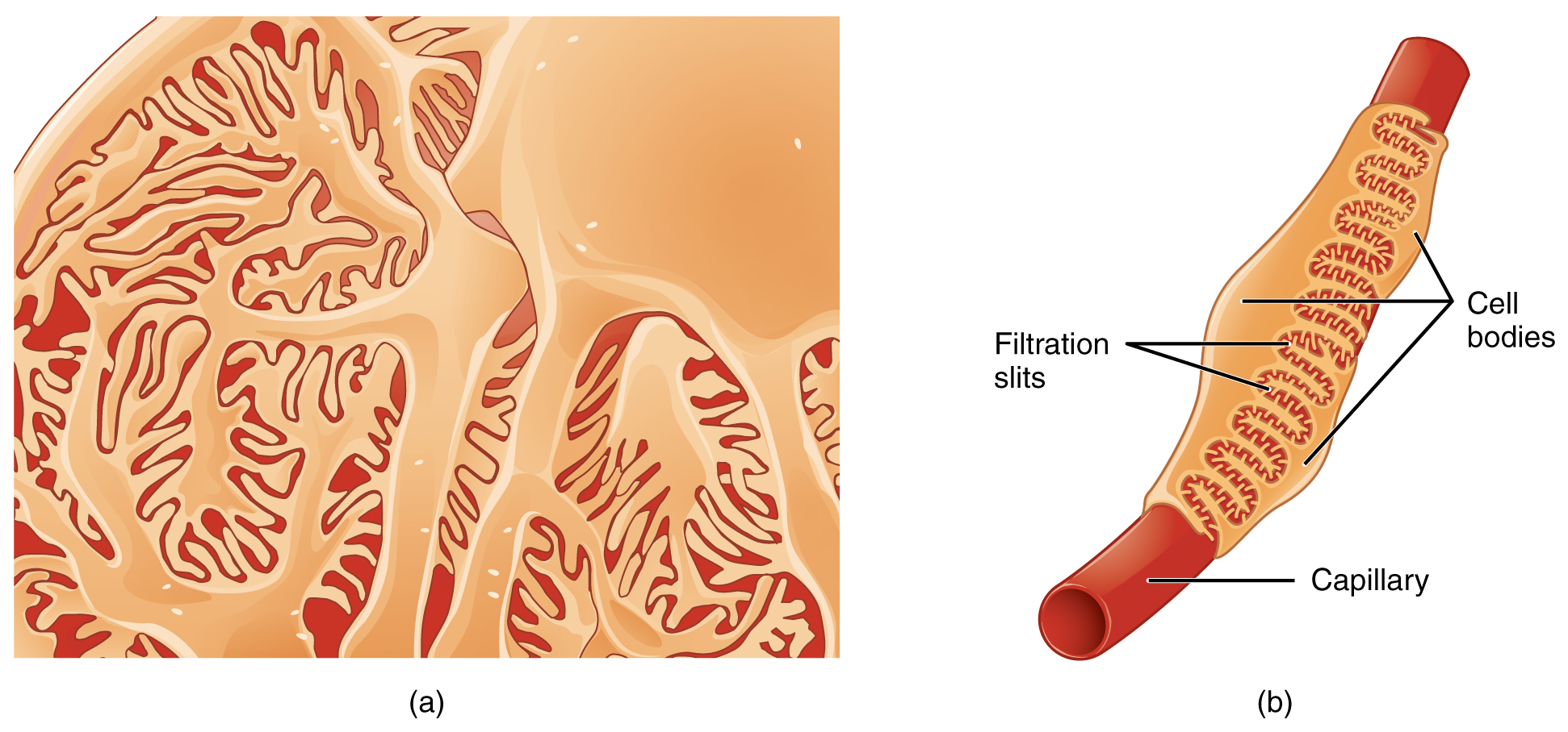
Filtration begins at the glomerulus, where blood pressure is high. Between the blood and the interior of the capsule lies the filtration membrane, which has three layers: the fenestrated endothelium of the glomerular capillaries, a basement membrane, and the podocytes of the capsule with gaps called filtration slits between their foot processes. This membrane acts as a selective barrier. Water and small solutes pass freely; blood cells are held back by the endothelium and the slits; and most plasma proteins are held back by the basement membrane. The result is a filtrate that resembles plasma in its concentration of ions, glucose and amino acids but contains almost no protein. The volume filtered each minute is the glomerular filtration rate, about 125 millilitres per minute, which adds up to roughly 180 litres a day.
For example: In glomerulonephritis, damage to the filtration membrane lets protein into the filtrate, so protein appears in the urine.
 Enlarge illustration ↗
Look at the interlocking foot processes and the narrow slits between them. This close-up shows the podocyte side of the filtration barrier; the capillary endothelium and basement membrane are additional layers, not replaced by these slits.
Image source and licence
OpenStax College · Source image · CC BY 3.0. Original image, reproduced without alteration.
Reabsorption: reclaiming what was filtered
Reabsorption returns most of the filtrate to the blood, and it happens mainly in the proximal convoluted tubule. There, cells actively transport sodium out of the tubular fluid, and other substances follow: glucose and amino acids are carried in with sodium, and water follows osmotically. This is why the proximal tubule reclaims the great majority of the filtered water, ions, glucose and amino acids. The loop of Henle and the distal convoluted tubule perform finer adjustments, and hormones act there to control how much water and salt are retained. Reabsorption is not unlimited: each carrier protein has a maximum rate, called the transport maximum. When the amount of a substance filtered exceeds that maximum, the excess cannot be reabsorbed and is lost in the urine, which is what happens to glucose in diabetes.
For example: Almost all the glucose filtered each day is reabsorbed; only when blood glucose is very high does the transport maximum fail and glucose reach the urine.
Secretion: adding what must be removed
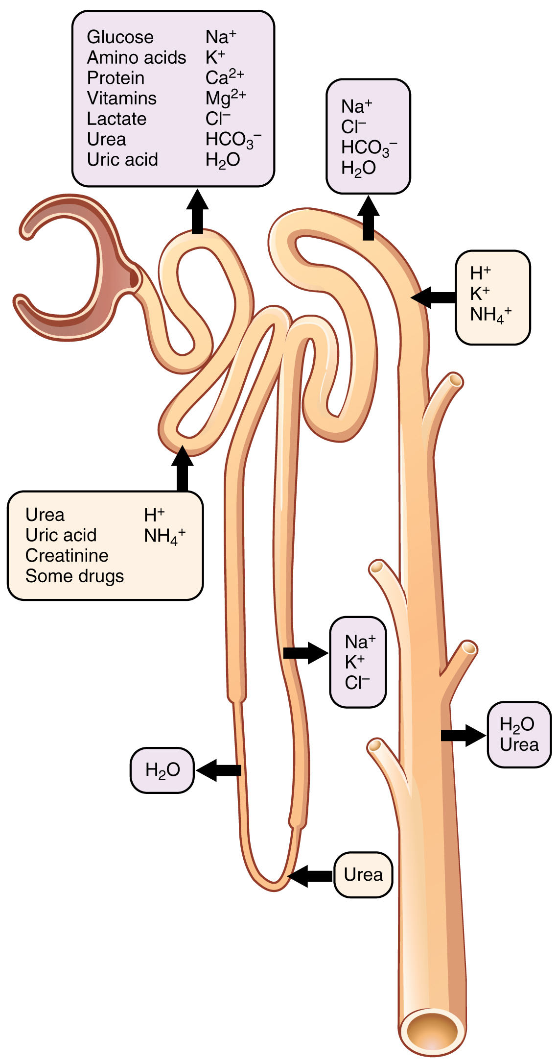
Secretion moves substances in the opposite direction to reabsorption: from the peritubular blood into the tubular fluid. It is important for two reasons. First, it removes waste products that filtration alone does not clear efficiently, including hydrogen ions, potassium, ammonium and creatinine, as well as many drugs and their breakdown products. Second, it gives the kidney fine control over blood composition, because secretion can continue even when filtration is reduced. Hydrogen ion secretion, for example, is central to the regulation of blood pH: by adding hydrogen ions to the urine, the kidney removes acid from the body. Potassium secretion in the distal tubule is controlled by aldosterone and is one of the ways the body keeps potassium within a narrow range.
For example: Penicillin is removed from the blood largely by tubular secretion, which is why some drugs are given with another medicine that blocks that secretion to prolong their effect.
 Enlarge illustration ↗
Read each arrow relative to the tubule. Outward arrows show reabsorption toward the surrounding tissue and blood; inward arrows show secretion into tubular fluid. Find the H⁺ arrows: adding hydrogen ions to the tubular fluid helps remove acid from the body.
Image source and licence
OpenStax College · Source image · CC BY 3.0. Original image, reproduced without alteration.
The loop of Henle and concentrated urine
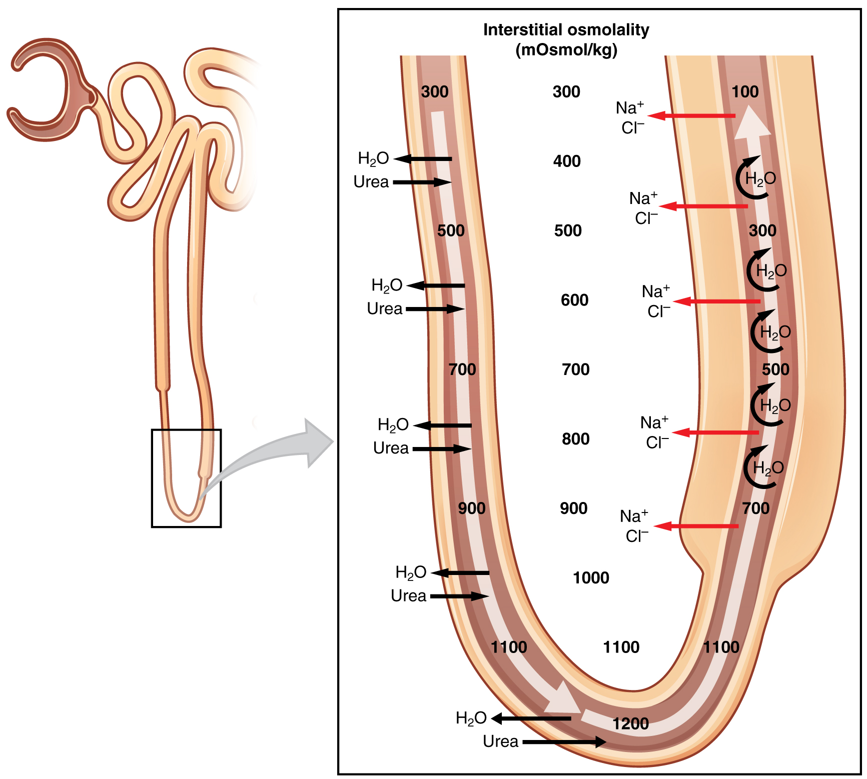
Concentrating urine requires a gradient, and the loop of Henle builds it. The descending limb is permeable to water but not very permeable to salt, so as filtrate descends into the salty medulla, water leaves and the fluid becomes more concentrated. The ascending limb is the opposite: it is impermeable to water but actively transports sodium and chloride out into the medullary tissue, which is what makes the medulla salty in the first place. Because the two limbs work in opposite directions and are close together, the arrangement is called a countercurrent multiplier. The collecting duct then passes through the same salty medulla, and if it is permeable to water, water is drawn out and the urine becomes concentrated. When the body needs to conserve water, antidiuretic hormone increases that permeability, so more water is reclaimed.
For example: After a long period without drinking, antidiuretic hormone rises, the collecting ducts become more permeable to water, and a small volume of concentrated urine is produced.
 Enlarge illustration ↗
Follow the descending limb down and the ascending limb up. Water can leave the descending limb; the ascending limb does not let water follow the salt leaving it. The numbers illustrate a concentration gradient, not a fixed value in every kidney.
Image source and licence
OpenStax College · Source image · CC BY 3.0. Original image, reproduced without alteration.
Key facts to remember
- Filtration occurs at the renal corpuscle, driven by blood pressure, and produces filtrate similar to plasma without most proteins.
- The filtration membrane has three layers: fenestrated endothelium, basement membrane and podocytes with filtration slits.
- Glomerular filtration rate is about 125 mL/min, roughly 180 litres a day, while urine output is about 1–2 litres a day.
- Reabsorption returns about 99% of the filtrate to the blood, mainly in the proximal convoluted tubule, and is limited by the transport maximum.
- Secretion moves hydrogen ions, potassium, ammonium, creatinine and drugs from the blood into the tubule; the countercurrent loop of Henle creates the medullary gradient that concentrates urine.
Example · Trace one pass through the nephron. About 125 mL of filtrate form each minute in the corpuscles. In the proximal tubule, sodium is pumped out and glucose, amino acids and water follow, reclaiming most of the fluid. In the loop of Henle, the descending limb loses water and the ascending limb loses salt, building the medullary gradient. In the distal tubule and collecting duct, hormones fine-tune sodium and water reabsorption, and hydrogen and potassium are secreted. What leaves as urine is roughly 1–2 litres a day containing urea, creatinine, excess ions and the products of secretion. Three processes, one product.
Read full notes
Read one topic at a time. These notes explain the mechanisms and relationships behind the key facts.
Three processes, one product
Describing urine formation as three processes is useful because each can be measured and each can fail independently. Filtration depends on renal blood flow and glomerular pressure; if either falls, less filtrate forms. Reabsorption depends on carrier proteins, energy supply and hormones; if it fails, useful substances are lost. Secretion depends on tubular cell function; if it fails, waste products and drugs accumulate. The final urine reflects the sum. This is why a single urine sample can reveal so much: glucose suggests the reabsorptive limit was exceeded, protein suggests the filtration barrier is damaged, and a very dilute urine suggests that water reabsorption is impaired.
Filtration and the filtration membrane
The filtration membrane explains what filtrate is and what it is not. Because the barrier has three layers of different sizes, it filters by both size and charge. Small molecules pass, cells do not, and most proteins are retained. This selectivity is why the presence of protein or blood in urine is abnormal and points to damage. The rate of filtration is also remarkable: about 125 millilitres a minute, or 180 litres a day, which is many times the total plasma volume. Such a large throughput is possible because the glomerular capillaries are highly permeable and the pressure within them is kept high. It also means that small changes in pressure produce large changes in filtrate volume, which is why the body regulates glomerular pressure carefully.
Reabsorption: reclaiming what was filtered
The scale of reabsorption is the key fact: about 99% of the filtrate is returned to the blood, and only 1–2 litres a day is excreted. This means the tubule is not a drain but a recovery system. The proximal tubule does most of the work, reclaiming water, ions, glucose, amino acids and small proteins. Later segments adjust the proportions under hormonal control, so the final urine can be dilute or concentrated, acidic or alkaline, rich or poor in potassium, according to the body's needs. Two features make this possible: carrier proteins that move substances against or with gradients, and the ability of the tubule to change its permeability in response to hormones such as ADH and aldosterone.
Secretion: adding what must be removed
Secretion is often overlooked because reabsorption is larger, but it is essential. Filtration alone would remove only what is small enough to pass the barrier and present in high enough concentration. Secretion allows the kidney to dispose of substances that are protein-bound and therefore poorly filtered, and to remove substances that must be eliminated quickly. It is also the mechanism by which the kidney regulates pH: hydrogen ions are secreted into the tubule to remove acid, and bicarbonate is reclaimed to keep the blood alkaline. Potassium secretion in the distal nephron, controlled by aldosterone, is another example. Many drugs are also secreted, which is why kidney function affects drug levels in the blood.
The loop of Henle and concentrating urine
The countercurrent arrangement is the reason mammals can produce urine more concentrated than their blood. The descending limb loses water and the ascending limb loses salt, so the medullary tissue becomes increasingly salty with depth, and the fluid entering the distal tubule is dilute. The collecting duct then passes through the salty medulla, and water can be drawn out if the duct is permeable. Because permeability is controlled by ADH, the same anatomy can produce either a large volume of dilute urine or a small volume of concentrated urine. This is a good example of how a structural arrangement and a hormonal signal combine to give the body flexible control over water balance.
Explore the diagram
Start with the whole view, then select a numbered structure. Compare the additional illustrations to connect what a structure looks like with what it does.
From blood to urine: nephron and tract
A schematic teaching view, not to scale: a small figure places the kidneys in the body, a frontal kidney section shows cortex, medulla and pelvis, an enlarged nephron shows where filtration, reabsorption and secretion happen, and the lower part of the figure shows the tract from ureter to urethra.
More controls
01 / 22 Body position reference
Blood arrives and is filtered under pressure
Body position reference
The kidneys lie high on the posterior abdominal wall, one on each side of the vertebral column. The right kidney usually sits slightly lower because the liver occupies space above it. They are retroperitoneal, meaning they lie behind the peritoneum rather than inside it.
Follow fluid through a nephron
One simplified nephron with its collecting duct; neighbouring nephrons are omitted. Blood and tubular fluid occupy separate spaces. The cortex is above the dashed boundary.
The glomerular capillaries lie inside a capsule. Filtration moves water and small solutes from blood into the capsular space; cells and most proteins remain in blood.
Connect the illustrations to the key ideas
FILTRATION · glomerulus to capsule, driven by pressure
Cells and most proteins are retained; filtrate resembles plasma without them.
REABSORPTION AND SECRETION · useful substances back to blood; wastes into the tubule
About 99% is reabsorbed; hydrogen, potassium, ammonium and drugs are secreted.
URINE · water, urea, creatinine, ions
The countercurrent loop and ADH set whether urine is dilute or concentrated.
Core Concepts Defined
- Glomerular filtration
- The pressure-driven movement of fluid from the glomerulus into the glomerular capsule.
- Filtrate
- The fluid entering the tubule, similar to plasma without most proteins.
- Filtration membrane
- The three-layered barrier between blood and the capsule: endothelium, basement membrane and podocytes.
- Fenestration
- A pore in the glomerular capillary endothelium.
- Basement membrane
- The middle layer of the filtration membrane, which holds back most plasma proteins.
- Podocyte
- A cell of the glomerular capsule whose foot processes form filtration slits.
- Filtration slit
- A gap between podocyte foot processes through which filtrate passes.
- Glomerular filtration rate (GFR)
- The volume filtered by both kidneys each minute, about 125 mL.
- Reabsorption
- Movement of substances from the tubular fluid back into the blood.
- Secretion
- Movement of substances from the blood into the tubular fluid.
- Transport maximum
- The maximum rate at which a carrier can move a substance during reabsorption.
- Renal threshold
- The plasma concentration above which a substance begins to appear in urine.
- Countercurrent multiplier
- The arrangement of the two limbs of the loop of Henle that builds the medullary solute gradient.
- Tubular fluid
- The fluid inside the tubule, which becomes urine as it is modified.
- Urea
- The main nitrogenous waste product in urine, formed from protein breakdown.
- Creatinine
- A waste product from muscle metabolism, filtered and secreted into urine.
- Antidiuretic hormone (ADH)
- A hormone that increases water permeability of the collecting ducts, concentrating urine.
Go deeper · worked example
Why glucose appears in urine in diabetes
- Filtration is passive and unlimited. Glucose is small and freely filtered at the glomerulus, so the amount entering the tubule depends mainly on its plasma concentration and the filtration rate.
- Reabsorption is carrier-mediated and limited. Glucose is reabsorbed in the proximal tubule by carrier proteins that move it together with sodium, and each carrier has a maximum rate.
- Compare filtered load with transport maximum. While the filtered load is below the transport maximum, all the glucose is reabsorbed and none appears in urine.
- Exceed the threshold. When plasma glucose rises enough that the filtered load exceeds the transport maximum, the carriers are saturated. The excess glucose cannot be reabsorbed.
- Glucose appears in urine. The unreabsorbed glucose remains in the tubular fluid and is excreted. Because glucose carries water with it osmotically, urine volume also increases, which is why high blood glucose causes frequent urination.
Explain it yourself · Explain why a person with normal blood glucose has no glucose in the urine, while a person with very high blood glucose does.
Reveal a model explanation
Glucose is freely filtered at the glomerulus, so the amount entering the tubule each minute is proportional to its concentration in plasma. In the proximal tubule, glucose is reabsorbed by carrier proteins that also move sodium, and these carriers have a maximum transport rate. When blood glucose is normal, the filtered load is well below that maximum, so all the filtered glucose is reabsorbed and none is excreted. When blood glucose rises above the renal threshold, the filtered load exceeds the transport maximum, the carriers are saturated, and the excess glucose stays in the tubular fluid and appears in the urine. Because glucose is osmotically active, it also holds water in the tubule, increasing urine volume.
Check
Answer three questions from this section — one easy, one medium and one hard. If you miss one, you get a fresh set with explanations for the last attempt.
Practise this sectionOpen all questions and filter by difficulty or Bloom levelReferences and review status
OpenStax, Anatomy and Physiology 2e, 25.5 Tubular Reabsorption, 25.6 Tubular Secretion, 25.7 Regulation of Renal Blood Flow and 25.8 Regulation of Urine Concentration; Tortora & Derrickson, Principles of Anatomy and Physiology, 12e, ch. 26. Author review only; medical and French-language expert review pending.
Original teaching schematics. English and French wording is author-reviewed for study; anatomy and French-language expert review is pending. Checks are practice only, not a graded examination.
Practice progress is saved on this device for the current learner.