SECTION 3.1
Epithelial and connective tissues
Distinguish epithelial from connective tissue by structure, location and function, and recognise their main types.
Learn
A tissue is a group of similar cells and the material around them, working together for a common function. The body has four tissue families, and this section compares the first two. Epithelial tissue covers and lines surfaces and forms glands; its cells are packed closely together with very little material between them. Connective tissue supports, binds, protects and transports; its cells are usually widely spaced, separated by a large amount of material called the matrix. That difference in arrangement explains almost everything else: epithelium forms barriers and exchange surfaces, while connective tissue fills spaces, joins structures and carries materials. Learning to tell them apart is the first step in reading any organ, because most organs contain both.
Key Concepts & Topics
What makes a tissue
A tissue is not simply a collection of identical cells. It includes the cells and the material they secrete around themselves, and the two together perform a function the cells could not perform alone. That surrounding material is the extracellular matrix, often shortened to matrix. The amount and composition of the matrix vary enormously. In epithelial tissue there is almost none, so the cells touch one another and form a continuous sheet. In connective tissue the matrix dominates, and the cells are scattered within it. The four tissue families are epithelial, connective, muscle and nervous tissue, and most organs contain several of them. Recognising which family dominates a region tells you what that region is likely to do: cover, support, contract or signal.
For example: The lining of your mouth is mostly epithelium with almost no matrix, so it forms a smooth barrier. The tissue beneath it is mostly connective tissue, with abundant matrix that gives the cheek its flexibility.
Epithelial tissue: covering, lining and secreting
Epithelial tissue forms sheets of closely packed cells that cover the body surface, line internal cavities and tubes, and form glands. Because the cells are tightly joined, epithelium is an effective barrier: it protects underlying tissue, limits water loss, and resists injury and infection. It also specialises in exchange: a thin epithelium allows substances to filter or diffuse across it, and a thicker one protects. Epithelial cells have two distinct sides. The apical surface faces the outside or the lumen, the space inside a tube; the basal surface rests on a thin supporting layer called the basement membrane, which anchors the epithelium to the tissue beneath. Epithelium has no blood vessels of its own, a property described as avascular, so it receives nutrients by diffusion from the connective tissue below.
For example: The epidermis of the skin is a stratified epithelium that resists abrasion, while the single layer lining the air sacs of the lungs is thin enough for gases to cross.
Common misconception: Avascular does not mean inactive. Epithelium is nourished by diffusion from the connective tissue beneath it and is often very metabolically busy.
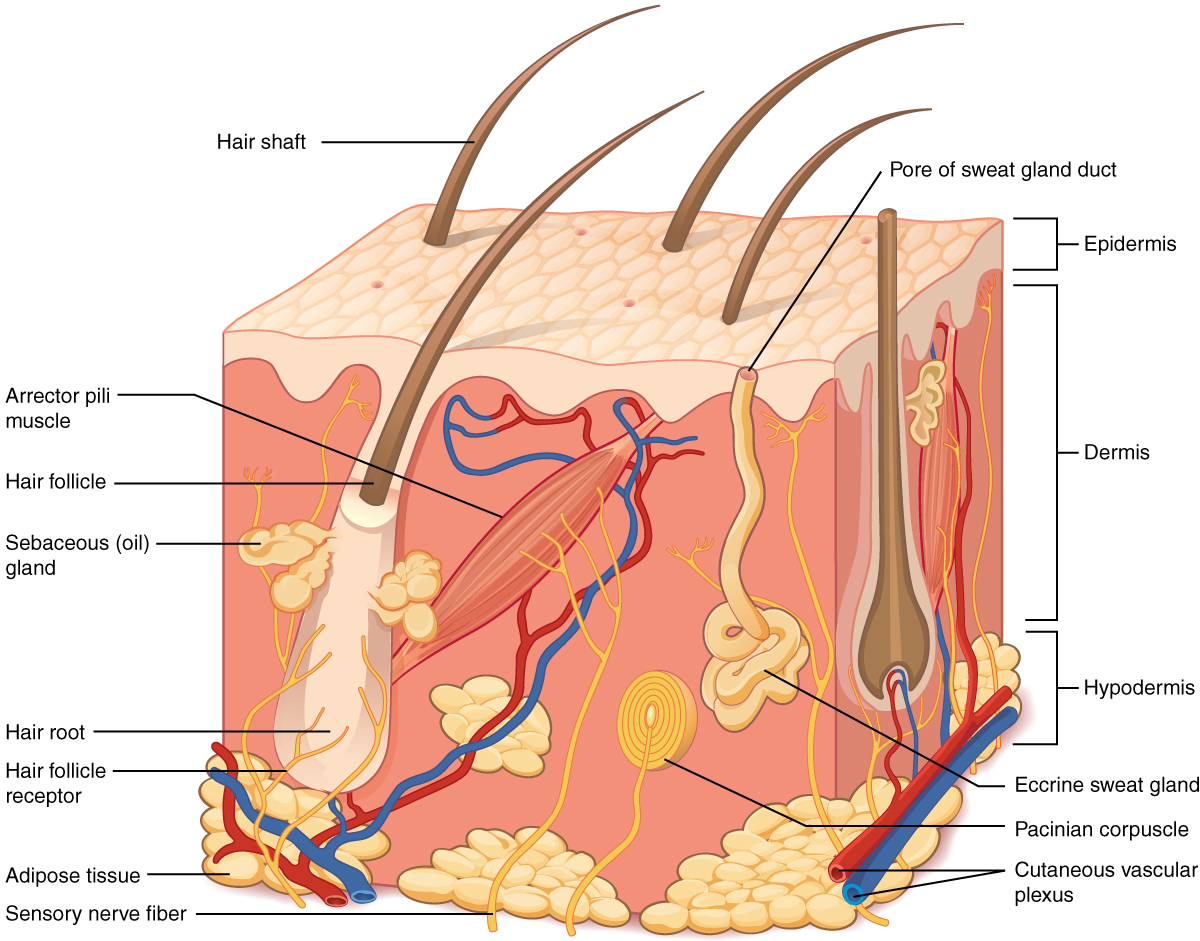 Enlarge illustration ↗
Find the thin surface epithelium above the deeper supporting tissue. Hair follicles and glands extend below the surface. This makes the difference between a surface lining and an entire organ easier to see.
Image source and licence
OpenStax College : J. Gordon Betts, Peter Desaix, Eddie Johnson. · Source image · CC BY 3.0. Original image, reproduced without alteration.
Simple and stratified epithelia
Epithelia are classified in two ways: by the number of cell layers and by the shape of the cells. A simple epithelium has one layer, which makes it thin and suited to exchange, absorption or secretion. A stratified epithelium has several layers, which makes it durable and suited to protection in places exposed to friction, such as the skin, mouth and oesophagus. Cell shape adds the second part of the name. Squamous cells are flat and thin, cuboidal cells are about as tall as they are wide, and columnar cells are taller than they are wide. Combining the two descriptions gives names such as simple squamous, simple columnar and stratified squamous. The name predicts both the appearance and the job: thin and flat for exchange, tall and layered for protection or secretion.
For example: Simple squamous epithelium lines the air sacs because it must be thin; stratified squamous epithelium lines the oesophagus because swallowed food scrapes the surface.
Connective tissue: cells in a matrix
Connective tissue is the most varied family in the body, and it is defined less by its cells than by its matrix. The matrix has two parts: ground substance, a gel that fills the space and holds water, and protein fibres. Collagen fibres are strong and resist stretching, elastic fibres stretch and recoil, and reticular fibres form fine supporting networks. The main cell type, the fibroblast, produces the matrix. Because the matrix can be soft, firm or hard, connective tissue ranges from loose packing material to firm cartilage to rigid bone, and it also includes blood, whose matrix is the fluid plasma. This variety explains its many roles: binding organs together, supporting and protecting them, insulating the body, storing energy and transporting substances.
For example: Loose connective tissue beneath the skin allows it to move over the tissues underneath, while dense connective tissue in a tendon transmits the pull of a muscle to bone.
Comparing epithelial and connective tissue
Four contrasts summarise the difference. Cell arrangement: epithelial cells are tightly packed and form sheets; connective cells are scattered in abundant matrix. Blood supply: epithelium is avascular and nourished by diffusion; most connective tissue is well supplied with blood, except cartilage. Position: epithelium covers surfaces and lines cavities and ducts; connective tissue fills spaces, binds structures and forms the body’s supporting framework. Main functions: epithelium protects, absorbs, filters and secretes; connective tissue supports, binds, protects, insulates, stores and transports. These contrasts are not rules to memorise blindly but consequences of the matrix. A tissue with almost no matrix must be a sheet; a tissue with a large matrix must be a supporting or transporting material.
For example: The gut wall has epithelium facing the food, connective tissue holding the layers together, muscle moving the contents and nerve tissue controlling it — four families in one organ.
Key facts to remember
- A tissue is similar cells plus the matrix around them, working together for a common function.
- Epithelial tissue forms sheets of tightly packed, avascular cells that cover, line, protect, absorb, filter and secrete.
- Epithelia are named by layers (simple or stratified) and cell shape (squamous, cuboidal or columnar).
- Connective tissue has widely spaced cells in an abundant matrix of ground substance and collagen, elastic or reticular fibres.
- The matrix decides whether connective tissue is loose, dense, adipose, cartilage, bone or blood.
Example · Look at the back of your hand. The surface is stratified squamous epithelium, avascular and protective. Beneath it lies loose connective tissue that allows the skin to move, then dense connective tissue that anchors it, and finally the bones and joints supported by connective tissue. The same small area contains several connective tissue types, all built on the same principle: cells scattered in a matrix whose composition sets the tissue’s job.
Read full notes
Read one topic at a time. These notes explain the mechanisms and relationships behind the key facts.
Four families, one principle
The four tissue families — epithelial, connective, muscle and nervous — are distinguished by the relationship between their cells and their matrix. Epithelium has cells packed tightly with almost no matrix, which suits covering and exchange. Connective tissue has a large matrix with scattered cells, which suits support, binding and transport. Muscle tissue has elongated cells full of contractile proteins, which suits producing force. Nervous tissue has cells specialised to generate and conduct electrical signals. In each case the arrangement of cells and matrix explains the function. When you study an organ, ask which families are present and which dominates in each layer; the answer usually explains both the organ’s appearance and its behaviour.
Epithelial tissue: form follows function
Every feature of epithelium serves its job. Tight junctions between cells stop material leaking between them, so the sheet is an effective barrier. The apical surface may carry tiny projections called microvilli that increase surface area for absorption, or cilia that move substances along. The basal surface rests on the basement membrane, which anchors the sheet and guides repair. The absence of blood vessels keeps the sheet thin, which is essential where exchange must be rapid, and nutrients reach the cells by diffusion from the connective tissue below. Because the cells divide frequently, most epithelia renew quickly, which is why they heal well but are also vulnerable to damage from radiation and some drugs that target dividing cells.
Simple and stratified epithelia
The choice between simple and stratified epithelium is a trade-off between thinness and durability. A simple epithelium is one cell thick, so substances cross it easily; this suits air sacs, capillaries and the lining of the gut, where exchange or absorption is the priority. A stratified epithelium has several layers, so the surface can be worn away and replaced without exposing the tissue beneath; this suits the skin, mouth, oesophagus and vagina, where friction is constant. Cell shape adds further specialisation: squamous cells give the thinnest possible barrier, cuboidal cells are common in glands and ducts, and columnar cells are found where absorption or secretion happens. Learning the naming system lets you predict both where an epithelium is found and what it does.
Connective tissue: cells in a matrix
Connective tissue is best understood from its matrix. Ground substance holds water and allows substances to diffuse, while fibres provide strength and elasticity. Collagen resists tension, so it is abundant in tendons, ligaments and the dermis. Elastic fibres allow tissues such as the lungs and large arteries to stretch and recoil. Reticular fibres form delicate networks that support soft organs such as lymph nodes and the spleen. By changing the proportions of these components, the same basic tissue becomes loose packing, strong tendon, flexible cartilage, rigid bone or fluid blood. The cells are relatively few, but they are not passive: fibroblasts build and maintain the matrix, and other connective tissue cells store fat, defend against microbes or remove debris. Connective tissue is therefore both a building material and a living system.
Comparing the two families in practice
In a real organ the two families work together. Epithelium forms the interface with the outside world or with a cavity, deciding what passes; connective tissue supports that interface, supplies it with blood and carries away what it produces. This partnership explains several clinical observations. An epithelium that is damaged heals quickly because it is supplied by the connective tissue beneath. A connective tissue disease can starve an epithelium of nutrients even when the epithelium itself is healthy. Wounds that reach only epithelium heal without a scar; wounds that damage connective tissue repair with collagen and leave a scar, because the matrix is not rebuilt exactly as before. Reading an organ as layers of different tissues makes these observations predictable rather than surprising.
Explore the diagram
Start with the whole view, then select a numbered structure. Compare the additional illustrations to connect what a structure looks like with what it does.
A lining and its supporting tissue
Enlarged section of a generic mucosa. Compare tightly packed epithelial cells with cells scattered in connective-tissue matrix.
Closely joined cells face a free surface or lumen. This example shows a single columnar layer; other epithelia have different cell shapes or several layers.
Connect the illustrations to the key ideas
EPITHELIAL TISSUE · tightly packed cells, no matrix
Forms sheets that cover, line and secrete; avascular and nourished by diffusion.
CONNECTIVE TISSUE · scattered cells in abundant matrix
The matrix of ground substance and fibres sets whether it is loose, dense, cartilage, bone or blood.
TOGETHER IN AN ORGAN · epithelium over connective tissue
The epithelium decides what passes; the connective tissue supports it and supplies blood.
Core Concepts Defined
- Tissue
- A group of similar cells and the matrix around them, working together for a common function.
- Extracellular matrix
- The material secreted around cells; its amount and composition give a tissue its properties.
- Epithelial tissue
- Sheets of tightly packed cells that cover surfaces, line cavities and ducts, and form glands.
- Apical surface
- The free surface of an epithelial cell, facing the exterior or the lumen.
- Basal surface
- The attached surface of an epithelial cell, resting on the basement membrane.
- Basement membrane
- A thin layer that anchors an epithelium to the connective tissue beneath it.
- Avascular
- Having no blood vessels of its own; epithelium is avascular and nourished by diffusion.
- Simple epithelium
- An epithelium of one cell layer, suited to exchange, absorption or secretion.
- Stratified epithelium
- An epithelium of several layers, suited to protection where friction occurs.
- Squamous
- Flat and thin, as in cells suited to exchange.
- Cuboidal
- About as tall as it is wide.
- Columnar
- Taller than it is wide, often with secretory or absorptive specialisations.
- Glandular epithelium
- Epithelium specialised to secrete products such as enzymes, sweat or hormones.
- Connective tissue
- Tissue with widely spaced cells in an abundant matrix that supports, binds, protects, stores and transports.
- Ground substance
- The gel part of the matrix that fills space and holds water.
- Collagen fibre
- A strong matrix fibre that resists stretching.
- Elastic fibre
- A matrix fibre that can stretch and recoil.
- Reticular fibre
- A fine matrix fibre that forms supporting networks.
- Fibroblast
- The main connective tissue cell, which produces the matrix.
- Loose connective tissue
- A soft packing tissue that binds organs and allows movement.
- Dense connective tissue
- A strong tissue with many collagen fibres, forming tendons and ligaments.
- Adipose tissue
- Connective tissue specialised for fat storage, insulation and cushioning.
- Cartilage
- A firm, flexible connective tissue, avascular, that cushions joints and supports passages.
- Bone
- A rigid connective tissue whose mineralised matrix supports and protects.
- Blood
- A connective tissue whose matrix is the fluid plasma and whose cells are the formed elements.
Go deeper · worked example
Why skin and the tissue under it behave differently
- Identify the surface layer. The outer layer of the skin is stratified squamous epithelium, with many layers of flat cells and almost no matrix.
- Link structure to function. Because the cells are tightly packed and layered, the epidermis resists abrasion, limits water loss and blocks many microbes. It has no blood vessels, so it depends on diffusion from below.
- Identify the layer beneath. The dermis is mostly connective tissue, with scattered cells in a matrix of collagen and elastic fibres, plus blood vessels, nerves and glands.
- Link structure to function. The abundant matrix lets the dermis stretch and recoil, its vessels nourish the epidermis and regulate temperature, and its fibres give the skin strength.
- Explain the consequence of each layer failing. If the epidermis is scraped, the barrier is lost and infection risk rises; if the dermis loses collagen or elastic fibres, the skin sags and tears more easily.
Explain it yourself · Explain why a shallow scrape of the skin bleeds very little, while a deeper cut bleeds freely.
Reveal a model explanation
The epidermis is epithelial tissue and has no blood vessels, so a scrape that removes only epidermis damages no vessels directly; any blood comes from the underlying dermis. A deeper cut reaches the dermis and its connective tissue, which is well supplied with blood vessels. Because epithelium is avascular and most connective tissue is vascular, the depth of the wound determines how much bleeding occurs.
Check
Answer three questions from this section — one easy, one medium and one hard. If you miss one, you get a fresh set with explanations for the last attempt.
Practise this sectionOpen all questions and filter by difficulty or Bloom levelReferences and review status
OpenStax, Anatomy and Physiology 2e, 4.1 Types of Tissues, 4.2 Epithelial Tissue and 4.3 Connective Tissue Supports and Protects; Tortora & Derrickson, Principles of Anatomy and Physiology, 12e, ch. 4. Author review only; medical and French-language expert review pending.
Original teaching schematics. English and French wording is author-reviewed for study; anatomy and French-language expert review is pending. Checks are practice only, not a graded examination.
Practice progress is saved on this device for the current learner.