SECTION 9.2
Digestion and absorption
Explain mechanical and chemical digestion and describe where and how the main nutrients are absorbed.
Learn
Digestion turns food into molecules small enough to cross the intestinal wall, and absorption moves those molecules into the blood or lymph. Two kinds of digestion work together. Mechanical digestion is physical: chewing, churning and segmentation break food into smaller pieces and mix it with secretions, which increases the surface available to enzymes. Chemical digestion is enzymatic: specific enzymes split large molecules into their building blocks by adding water, a reaction called hydrolysis. Carbohydrates become monosaccharides, proteins become amino acids and fats become fatty acids and monoglycerides. These products are then absorbed in the small intestine, most of them into blood capillaries and the products of fat digestion into lymph. This section explains how each nutrient class is digested and where absorption occurs.
Key Concepts & Topics
Mechanical and chemical digestion
Mechanical digestion prepares food for chemical digestion. Chewing in the mouth reduces particle size; churning in the stomach mixes food with acid and enzymes; and segmentation in the small intestine repeatedly divides and recombines the contents so that they contact the absorbing surface. None of these processes changes the chemical composition of food, but all of them expose more surface to enzymes, which is why they matter. Chemical digestion is carried out by enzymes, and each enzyme acts on a specific type of molecule: amylases act on starches, proteases on proteins and lipases on fats. The reaction they catalyse is hydrolysis, in which a molecule of water is inserted to split a bond. This is why digestion requires water, and why the products of digestion are smaller and more soluble than the food that entered the tract.
For example: Chewing a piece of bread thoroughly makes it taste slightly sweet, because salivary amylase has begun to break starch into sugars.
Carbohydrate digestion and absorption
Carbohydrate digestion begins in the mouth, where salivary amylase starts breaking starch into shorter chains. Salivary amylase is inactivated by stomach acid, so most starch digestion resumes in the duodenum, where pancreatic amylase continues the work. The final step occurs at the brush border, the surface of the intestinal cells, where enzymes such as maltase, sucrase and lactase split disaccharides into monosaccharides. Only monosaccharides can be absorbed. Glucose and galactose are absorbed by active cotransport with sodium, while fructose is absorbed by facilitated diffusion. The absorbed monosaccharides enter the blood capillaries inside the villi and are carried by the hepatic portal vein to the liver, which regulates how much glucose circulates. This is why a person who lacks lactase cannot digest lactose and develops symptoms after drinking milk.
For example: Lactose intolerance causes bloating and diarrhoea because undigested lactose remains in the intestine, where bacteria ferment it and it draws in water.
 Enlarge illustration ↗
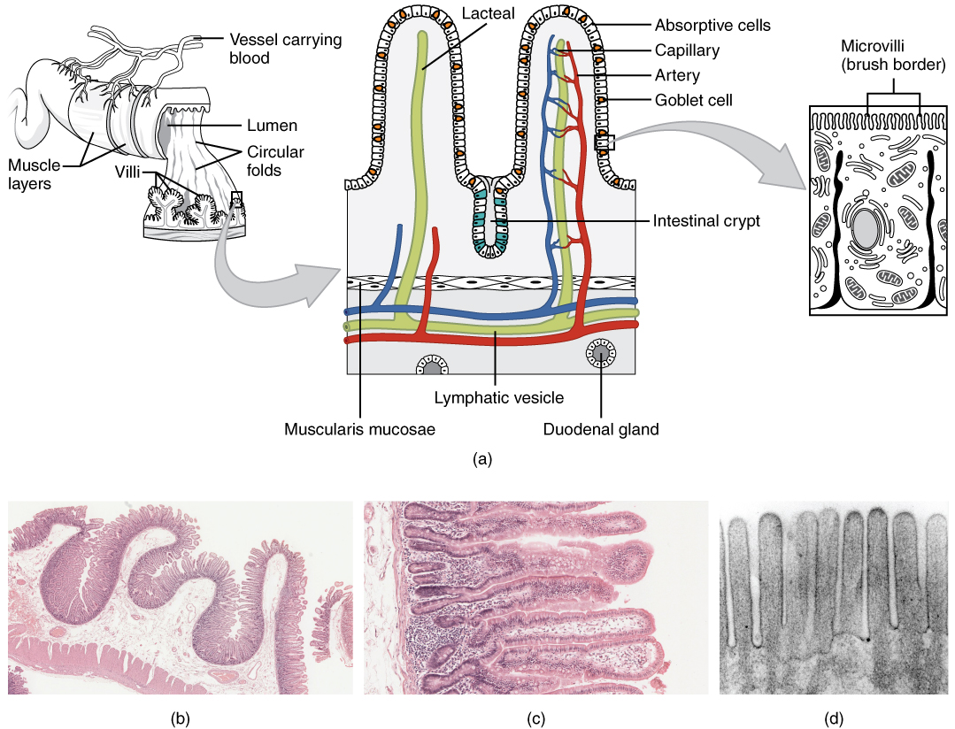
Move from the intestinal wall to a single villus and its epithelial surface. Folds, villi and microvilli are different levels of surface enlargement. They increase the area available for absorption.
Image source and licence
OpenStax College · Source image · CC BY 3.0. Original image, reproduced without alteration.
Protein digestion and absorption
Protein digestion starts in the stomach, where hydrochloric acid denatures proteins, unfolding them so that enzymes can reach their bonds, and pepsin begins splitting them into shorter peptides. Pepsin works only in acid, which is why it is active in the stomach and inactive once it reaches the alkaline duodenum. In the duodenum, pancreatic proteases continue the work: trypsin and chymotrypsin split peptides into smaller fragments, and carboxypeptidase removes amino acids from one end. These enzymes are released as inactive precursors and activated in the intestine, so they do not digest the pancreas itself. The final step takes place at the brush border, where peptidases split short peptides into single amino acids. Amino acids are absorbed by active cotransport with sodium, and like monosaccharides they enter the blood capillaries and travel to the liver by the hepatic portal vein.
For example: Because pepsin needs acid, drugs that strongly suppress stomach acid can reduce protein digestion in the stomach, though pancreatic proteases largely compensate.
Fat digestion and absorption
Fat digestion faces a problem: fats do not mix with water, so enzymes dissolved in the watery contents of the intestine cannot easily reach them. Bile salts solve this by emulsifying fats, breaking large droplets into many small ones suspended in the fluid. This does not digest the fat chemically; it simply gives pancreatic lipase a much larger surface to work on. Lipase then splits each triglyceride into a monoglyceride and two fatty acids. These products, together with bile salts, form tiny clusters called micelles that ferry the fats to the brush border, where they are absorbed by diffusion. Inside the intestinal cells, the products are re-assembled into triglycerides and packaged with proteins into chylomicrons. Because chylomicrons are too large for blood capillaries, they enter the lacteals, the lymphatic capillaries of the villi, and travel in lymph before reaching the blood.
For example: After a fatty meal, lymph from the intestine looks milky because it is carrying chylomicrons.
Common misconception: Bile does not digest fat. It emulsifies it, and the actual chemical digestion is carried out by pancreatic lipase.
Water, vitamins and minerals
Water is absorbed along the whole length of the intestine, mostly by osmosis in the small intestine, following the movement of solutes such as sodium and glucose. The large intestine absorbs the remaining water and electrolytes, which is why its failure causes watery diarrhoea. Vitamins follow different routes depending on their solubility. Fat-soluble vitamins A, D, E and K dissolve in the fat droplets and are absorbed with the products of fat digestion, so a disorder that impairs fat absorption also impairs their absorption. Most water-soluble vitamins are absorbed by specific carriers. Vitamin B12 is a special case: it must first bind to a protein called intrinsic factor, produced by the stomach, and the complex is then absorbed in the ileum. Minerals such as iron and calcium are absorbed by regulated carriers whose activity adjusts to the body's needs.
For example: Loss of intrinsic factor causes pernicious anaemia, because vitamin B12 cannot be absorbed even when the diet contains enough of it.
Key facts to remember
- Mechanical digestion breaks and mixes food without changing its chemistry; chemical digestion uses enzymes to split molecules by hydrolysis.
- Starch digestion uses salivary and pancreatic amylase, then brush-border maltase, sucrase and lactase; only monosaccharides are absorbed.
- Protein digestion uses acid and pepsin in the stomach, then pancreatic trypsin, chymotrypsin and carboxypeptidase, then brush-border peptidases.
- Fat is emulsified by bile salts and digested by pancreatic lipase into monoglycerides and fatty acids, absorbed as micelles and exported as chylomicrons in lymph.
- Water, fat-soluble vitamins, water-soluble vitamins and minerals are absorbed by different routes; vitamin B12 needs intrinsic factor and is absorbed in the ileum.
Example · Follow a mixed meal. Starch is partly digested in the mouth and further in the duodenum, then split at the brush border into glucose, which enters the blood and travels to the liver. Proteins are unfolded and partly split in the stomach, then digested by pancreatic proteases and brush-border peptidases into amino acids, which also enter the blood. Fats are emulsified by bile salts and digested by lipase; their products are absorbed as micelles, rebuilt into triglycerides, packaged into chylomicrons and carried away in lymph. Water follows solutes across the intestinal wall, fat-soluble vitamins travel with fats, and vitamin B12 travels with intrinsic factor to the ileum.
Read full notes
Read one topic at a time. These notes explain the mechanisms and relationships behind the key facts.
Mechanical and chemical digestion
The two types of digestion are partners rather than alternatives. Mechanical digestion without chemical digestion would leave food small but unusable, and chemical digestion without mechanical digestion would be slow because enzymes act only on surfaces. Together they reduce food to molecules that can be absorbed. Another feature of the system is the staging of enzymes: each enzyme works in the compartment where conditions suit it. Salivary amylase acts in the neutral mouth; pepsin acts only in the acid stomach; pancreatic enzymes act in the alkaline duodenum, protected by bicarbonate. This staging prevents enzymes from digesting the organs that produce them and ensures that each nutrient class is broken down at the right point in the tract.
Carbohydrate digestion and absorption
Carbohydrate digestion is a good illustration of a stepwise process. Starch is too large to be absorbed, so it is broken down progressively: amylase produces shorter chains, and brush-border enzymes produce single sugars. The final enzymes are attached to the intestinal surface rather than secreted into the lumen, which means the last stage of digestion happens exactly where absorption occurs. Glucose and galactose are taken up actively with sodium, so their absorption depends on the sodium gradient maintained by the cells; fructose uses a different, passive route. Once absorbed, the sugars enter the blood and pass to the liver, which buffers blood glucose and stores the excess as glycogen. Lactase deficiency shows what happens when one of these steps fails: undigested lactose remains in the lumen, draws in water osmotically and is fermented by bacteria.
Protein digestion and absorption
Protein digestion shows how the body protects itself from its own enzymes. Proteases are powerful and would digest the pancreas if released in active form, so they are secreted as inactive precursors and activated only in the intestinal lumen. Pepsin is likewise produced as pepsinogen and activated by acid. The stomach's acid has a second role: by unfolding proteins it exposes the peptide bonds that enzymes must reach, a process called denaturation. The final products, amino acids, are absorbed actively and travel to the liver, which uses them to build plasma proteins and to redistribute them to other tissues. Because the body cannot store surplus amino acids as a reserve, they are either used or broken down, and the nitrogen is excreted as urea by the kidneys.
Fat digestion and absorption
Fat digestion is a lesson in the importance of physical chemistry. Because fats are insoluble, the body cannot rely on enzymes meeting their substrate by simple diffusion; it must first emulsify the fat so that lipase has a workable surface. Bile salts perform this task and also form micelles that carry the digestion products through the watery layer next to the brush border. Inside the intestinal cell, the products are re-assembled, because triglycerides cannot cross the basolateral membrane easily; packaging them into chylomicrons solves both the transport and the size problem. The chylomicrons then enter lymph rather than blood, which is why the route of absorbed fat differs from that of sugars and amino acids and why disorders of lymph drainage can affect fat absorption.
Water, vitamins and minerals
The absorption of water, vitamins and minerals is easy to overlook but clinically important. Water absorption depends on solute absorption, because water follows osmotic gradients; this is why rehydration solutions contain both salt and glucose, which pull water into the body. Fat-soluble vitamins depend on fat absorption, so conditions that reduce fat absorption also cause deficiencies of vitamins A, D, E and K. Vitamin B12 depends on intrinsic factor and on a healthy ileum, so its deficiency can result from stomach disease, ileal disease or a vegetarian diet without supplementation. Minerals are absorbed by regulated transporters that respond to body needs, which is why iron absorption increases during iron deficiency. Each of these routes can fail independently.
Explore the diagram
Start with the whole view, then select a numbered structure. Compare the additional illustrations to connect what a structure looks like with what it does.
The digestive route
Trace the food pathway, then distinguish the accessory organs that add secretions.
More controls
01 / 7 Oesophagus
Oesophagus
A muscular tube carries swallowed material to the stomach.
Illustration credits
- Mariana Ruiz Villarreal (LadyofHats), Jmarchn · Digestive system without labels.svg · Public domain.

Adapted for this lesson: labels removed, artwork cropped or recoloured, and interactive teaching overlays added. Adapted artwork retains its source licence.
Two routes out of an intestinal villus
An enlarged villus projects into the intestinal lumen. Microvilli are much smaller projections on its epithelial cells, not additional whole villi.
A thin epithelial layer separates digested nutrients in the lumen from vessels inside the villus. Its microvilli increase the absorptive surface.
Connect the illustrations to the key ideas
MECHANICAL AND CHEMICAL · chewing, churning, segmentation; enzymes and hydrolysis
Mechanical work increases surface; enzymes split bonds by adding water.
NUTRIENT-SPECIFIC DIGESTION · amylases; proteases; lipase with bile salts
Starch to monosaccharides, protein to amino acids, fat to monoglycerides and fatty acids.
TWO ABSORPTION ROUTES · sugars and amino acids to blood; fats to lymph
Blood capillaries carry nutrients to the liver; lacteals carry chylomicrons.
Core Concepts Defined
- Mechanical digestion
- Physical breakdown and mixing of food by chewing, churning and segmentation.
- Chemical digestion
- Enzymatic splitting of food molecules into smaller absorbable units.
- Hydrolysis
- A reaction that splits a bond by adding a molecule of water.
- Enzyme specificity
- The property that each enzyme acts on one type of substrate.
- Amylase
- An enzyme that digests starch into shorter chains; found in saliva and pancreatic juice.
- Brush border
- The microvilli-covered surface of intestinal cells where final digestion occurs.
- Maltase, sucrase and lactase
- Brush-border enzymes that split disaccharides into monosaccharides.
- Pepsin
- A stomach enzyme that begins protein digestion and works only in acid.
- Trypsin and chymotrypsin
- Pancreatic proteases that split peptides into smaller fragments.
- Carboxypeptidase
- A pancreatic protease that removes amino acids from the end of a peptide.
- Emulsification
- The physical breaking of large fat droplets into small ones by bile salts.
- Micelle
- A small cluster of bile salts and fat-digestion products that ferries fats to the brush border.
- Chylomicron
- A large lipoprotein particle that carries absorbed fats into lymph.
- Lacteal
- The lymphatic capillary inside a villus that receives chylomicrons.
- Hepatic portal vein
- The vein carrying absorbed nutrients from the intestine to the liver.
- Intrinsic factor
- A stomach protein required for vitamin B12 absorption in the ileum.
- Monosaccharide
- A single sugar unit, the absorbable product of carbohydrate digestion.
Go deeper · worked example
How a mixed meal is digested and absorbed
- Mechanical preparation. Chewing reduces food to a bolus and mixes it with saliva, whose amylase begins starch digestion; stomach churning mixes it with acid and enzymes.
- Carbohydrates. Starch is split by pancreatic amylase in the duodenum and then by brush-border maltase and sucrase into monosaccharides, which are absorbed with sodium into blood capillaries.
- Proteins. Acid unfolds proteins and pepsin begins splitting them; pancreatic trypsin, chymotrypsin and carboxypeptidase continue, and brush-border peptidases release amino acids, absorbed into blood.
- Fats. Bile salts emulsify the fat, pancreatic lipase splits triglycerides into monoglycerides and fatty acids, micelles deliver them to the brush border, and the cells rebuild triglycerides and export chylomicrons into lacteals.
- Water, vitamins and minerals. Water follows solutes across the intestinal wall, fat-soluble vitamins travel with fats, and vitamin B12 travels bound to intrinsic factor to be absorbed in the ileum; the large intestine absorbs the remaining water.
Explain it yourself · Explain why fats are absorbed into lymph while sugars and amino acids are absorbed into blood.
Reveal a model explanation
Sugars and amino acids are small, water-soluble molecules. They cross the intestinal cells and pass directly into the blood capillaries inside each villus, from which they travel by the hepatic portal vein to the liver. Fats behave differently because they are not water-soluble. After absorption, the intestinal cells re-assemble the products into triglycerides and package them with proteins into large particles called chylomicrons. These particles are too large to enter blood capillaries, but the villi contain a lymphatic capillary, the lacteal, whose openings are wide enough to admit them. The chylomicrons therefore enter lymph, travel through lymphatic vessels and ducts, and reach the blood at the venous angles. This route also explains why fatty lymph looks milky after a fatty meal.
Check
Answer three questions from this section — one easy, one medium and one hard. If you miss one, you get a fresh set with explanations for the last attempt.
Practise this sectionOpen all questions and filter by difficulty or Bloom levelReferences and review status
OpenStax, Anatomy and Physiology 2e, 23.7 Chemical Digestion and Absorption: A Closer Look and 23.2 Digestive System Processes and Regulation; Tortora & Derrickson, Principles of Anatomy and Physiology, 12e, ch. 24. Author review only; medical and French-language expert review pending.
Original teaching schematics. English and French wording is author-reviewed for study; anatomy and French-language expert review is pending. Checks are practice only, not a graded examination.
Practice progress is saved on this device for the current learner.