SECTION 3.3
Body membranes and neuron structure
Identify the epithelial and connective-tissue membranes of the body and the main parts of a neuron.
Learn
The word membrane has two meanings in anatomy, and confusing them causes unnecessary difficulty. A cell membrane, also called the plasma membrane, is the thin boundary of a single cell, studied in section 2.1. A body membrane is a sheet of tissue that covers a surface, lines a cavity or separates structures. This section deals with body membranes and with the structure of the neuron. Body membranes are built from epithelium, connective tissue or both, and each type is suited to its location. The neuron is the signalling cell of nervous tissue, and its parts — cell body, dendrites and axon — are arranged so that signals travel in one direction with high speed. Learning both gives you the vocabulary to describe how the body covers, protects, lubricates and communicates.
Key Concepts & Topics
Two meanings of membrane
A cell membrane belongs to one cell. It is a phospholipid bilayer with proteins that controls what enters and leaves, and it is measured in nanometres. A body membrane is a macroscopic sheet of tissue, often several cell layers and their matrix, and it covers an organ, lines a cavity or forms a partition. The two are related — every body membrane is made of cells, and every cell has a cell membrane — but they answer different questions. When a clinician speaks of the mucous membrane of the nose, they mean a sheet of epithelium and connective tissue, not the boundary of a single cell. Keeping the two meanings separate prevents confusion when you read or write about membranes, and it makes the names of the body membranes easier to place.
For example: The cell membrane of one epithelial cell is invisible without a microscope. The mucous membrane lining the mouth is a visible sheet that you can touch and that renews itself continuously.
Common misconception: In anatomy, “membrane” does not always mean the boundary of a cell. Most named body membranes are sheets of tissue made of many cells.
Epithelial membranes
Epithelial membranes consist of an epithelium attached to an underlying layer of connective tissue, and there are three kinds. A mucous membrane, or mucosa, lines body passages that open to the exterior, such as the mouth, nose, digestive tract, airways and reproductive passages. It secretes mucus, which keeps the surface moist and traps particles. A serous membrane lines the closed cavities of the body and covers the organs inside them; it has a parietal layer on the cavity wall and a visceral layer on the organ, with lubricating serous fluid between them, as described in section 1.6. The pleura, pericardium and peritoneum are examples. The cutaneous membrane is the skin, which is exposed to the outside world; its epithelium is stratified and dry, and it protects the body rather than secreting mucus.
For example: The inside of your cheek is a mucous membrane: wet, renewing quickly and able to heal fast. The pleura around your lungs is a serous membrane: thin, lubricated and gliding with every breath.
 Enlarge illustration ↗
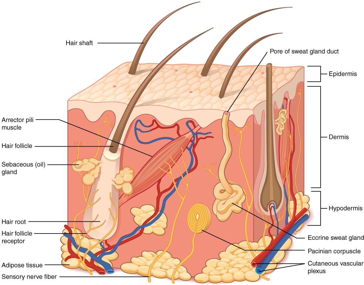
Find the thin surface epithelium above the deeper supporting tissue. Hair follicles and glands extend below the surface. This makes the difference between a surface lining and an entire organ easier to see.
Image source and licence
OpenStax College : J. Gordon Betts, Peter Desaix, Eddie Johnson. · Source image · CC BY 3.0. Original image, reproduced without alteration.
Connective-tissue membranes
Some membranes are made mainly of connective tissue rather than epithelium. A synovial membrane lines the cavity of a freely movable joint. It secretes synovial fluid, a slippery liquid that lubricates the joint surfaces, nourishes the cartilage and absorbs shock. Unlike epithelial membranes, it does not have a continuous epithelium covering its surface. The meninges are three connective-tissue membranes that cover the brain and spinal cord and enclose the cerebrospinal fluid. They protect the central nervous system, carry its blood supply in part and separate it from the bones around it. Fascia is a further connective-tissue sheet that wraps muscles, organs and nerves and holds them together. These membranes do not secrete mucus or serous fluid; their job is mechanical support, protection and lubrication of a different kind.
For example: A knee joint contains synovial fluid so the surfaces glide without friction; when the synovial membrane is inflamed, the joint becomes painful and swollen.
The parts of a neuron
A neuron has three main parts. The cell body, also called the soma, contains the nucleus and most of the cell’s organelles; it is the neuron’s control centre. Dendrites are short, branched extensions that receive signals from other neurons and carry them towards the cell body. The axon is a single long extension that carries the signal away from the cell body towards its target; it may be wrapped in a fatty myelin sheath that speeds conduction, with gaps called nodes of Ranvier between the wrapped segments. At its end, the axon branches into terminals that release chemical messengers onto the next cell. Because dendrites receive and the axon transmits, signals normally travel in one direction: dendrite → cell body → axon → terminal. This one-way arrangement is what allows nervous tissue to form reliable circuits.
For example: In a motor neuron, dendrites receive commands from the spinal cord, the cell body integrates them, and the axon carries the final signal all the way to a muscle fibre.
Types of neurons
Neurons are classified by the direction in which they carry information. Sensory neurons, also called afferent neurons, carry signals from receptors towards the central nervous system; they tell the brain and spinal cord what is happening. Motor neurons, or efferent neurons, carry signals from the central nervous system to muscles and glands; they produce action. Interneurons, or association neurons, lie within the central nervous system and connect sensory to motor pathways, allowing processing, comparison and coordination. This three-part arrangement explains the structure of a reflex: a sensory neuron detects the stimulus, an interneuron in the spinal cord processes it, and a motor neuron produces the response. Neurons can also be described by shape, but the functional classification is the more useful one in clinical practice because it predicts what a damaged neuron will affect.
For example: Touching a hot pan triggers a sensory neuron, an interneuron in the spinal cord and a motor neuron that pulls the hand away — all before the brain has consciously felt the pain.
Key facts to remember
- A cell membrane is the boundary of one cell; a body membrane is a sheet of tissue that covers, lines or separates.
- Epithelial membranes are mucous (lining passages open to the exterior), serous (lining closed cavities) and cutaneous (the skin).
- Synovial membranes line joint cavities and secrete synovial fluid; the meninges cover the brain and spinal cord.
- A neuron has a cell body with the nucleus, dendrites that receive signals and usually one axon that carries signals away.
- Sensory neurons carry signals inward, motor neurons carry them outward, and interneurons connect the two within the central nervous system.
Example · You step on a sharp object. Sensory neurons in the foot carry the signal to the spinal cord, where interneurons connect to motor neurons. The motor neurons send a signal back to the leg muscles, which contract and lift the foot. A moment later the brain registers the pain. In the same body, synovial membranes keep the knee joint lubricated, mucous membranes line the airways, serous membranes let the lungs glide, and the meninges protect the spinal cord through which the signal just travelled.
Read full notes
Read one topic at a time. These notes explain the mechanisms and relationships behind the key facts.
Two meanings of membrane
Most confusion about membranes comes from using one word for two scales. At the cellular scale, a membrane is a lipid bilayer a few nanometres thick that surrounds a cell or an organelle. At the tissue scale, a membrane is a sheet you can see and dissect: it may be several cell layers thick and include connective tissue. Textbooks usually signal the meaning by the context or by an adjective: plasma membrane and nuclear membrane are cellular, while mucous membrane, serous membrane and synovial membrane are tissue sheets. When you meet the word, ask what scale is being discussed. This small habit prevents a surprising number of mistakes, especially when a disease affects a tissue membrane while the cell membranes remain healthy, or the reverse.
Epithelial membranes: mucous, serous and cutaneous
The three epithelial membranes differ by where they face and what they secrete. A mucous membrane faces a passage that opens to the exterior, so it must stay moist and handle particles, microbes and food; it secretes mucus and often has a renewing epithelium and glands beneath. A serous membrane faces a closed cavity, so it must allow movement without friction; it secretes a thin serous fluid and has two layers that slide over one another. The cutaneous membrane faces the outside world directly, so it must be tough and relatively dry; its stratified epithelium resists abrasion and limits water loss. Each membrane also contains connective tissue beneath the epithelium, which supplies blood and supports repair — a reminder that tissues rarely work alone.
Connective-tissue membranes: synovial and meninges
Some membranes are connective tissue through and through. A synovial membrane lines the cavity of a freely movable joint and produces synovial fluid, a viscous liquid containing substances that nourish cartilage and reduce friction. Because cartilage has no blood vessels, it depends on this fluid for nutrients, which is why joint movement is good for cartilage health. The meninges are three layers around the brain and spinal cord. The outermost is tough and attached to the skull or vertebrae; the middle is filled with fluid and vessels; the innermost follows the surface of the nervous tissue. Between two of the layers lies cerebrospinal fluid, which cushions the brain, and the meninges themselves carry the blood supply and form a barrier against infection. These membranes show that connective tissue is not only a passive support: it can secrete, cushion, nourish and defend.
The parts of a neuron and the direction of the signal
The neuron’s shape is its function. Dendrites give a large surface for receiving signals from many sources, so the cell body can compare and integrate them. A single long axon lets one cell deliver a signal over a considerable distance, which is essential in a body measured in metres. Myelin around the axon increases speed by allowing the signal to be regenerated only at the nodes, rather than continuously along the whole membrane. Terminal branches spread the output to several targets at once. Every part has a clear role, and the arrangement enforces a single direction of travel: receiving surfaces on the dendrites and cell body, transmitting surface on the axon. This is why damage to different parts of a neuron produces different effects on sensation, movement or coordination.
Types of neurons and their jobs
Classifying neurons by direction of travel is clinically useful. Sensory neurons bring information in, so damage causes loss or alteration of sensation, or of the body’s awareness of its own position. Motor neurons carry commands out, so damage causes weakness or paralysis and reduced glandular secretion. Interneurons connect the two inside the central nervous system, so damage there can disrupt reflexes, coordination and the interpretation of sensory input without affecting the peripheral nerves themselves. A single clinical problem can involve more than one class: a spinal cord injury may interrupt sensory, motor and interneuronal pathways at once. Knowing the classes helps you predict the pattern of deficits a patient will show and which functions to test.
Explore the diagram
Start with the whole view, then select a numbered structure. Compare the additional illustrations to connect what a structure looks like with what it does.
A nerve pathway and a neuron
The body view locates the nervous system; the enlarged neuron explains how a cell receives and passes on a signal.
More controls
01 / 7 Brain
Brain
The brain is part of the central nervous system and integrates information.
Illustration credits
- Mariana Ruiz Villarreal (LadyofHats) · Complete neuron cell diagram en.svg · Public domain.

Adapted for this lesson: labels removed, artwork cropped or recoloured, and interactive teaching overlays added. Adapted artwork retains its source licence.
Skin as a living barrier
A magnified section shows layers, glands, vessels and sensory branches.
More controls
01 / 7 Epidermis
Epidermis
The outer epithelial layer forms a protective barrier and renews from deeper cells. It has no blood vessels.
Illustration credits
- Tomáš Kebert & umimeto.org · Human skin structure.svg · CC BY-SA 4.0.

Adapted for this lesson: labels removed, artwork cropped or recoloured, and interactive teaching overlays added. Adapted artwork retains its source licence.
A lining and its supporting tissue
Enlarged section of a generic mucosa. Compare tightly packed epithelial cells with cells scattered in connective-tissue matrix.
Closely joined cells face a free surface or lumen. This example shows a single columnar layer; other epithelia have different cell shapes or several layers.
Connect the illustrations to the key ideas
BODY MEMBRANES · mucous, serous, cutaneous, synovial, meninges
Each membrane is a tissue sheet suited to covering, lining, lubricating or protecting.
NEURON PARTS · cell body, dendrites, axon, terminals
Dendrites receive, the cell body integrates, the axon transmits and the terminals release messengers.
NEURON TYPES · sensory → interneuron → motor
Signals travel inward, are processed centrally and travel outward to muscles and glands.
Core Concepts Defined
- Cell (plasma) membrane
- The boundary of a single cell, made of a phospholipid bilayer with proteins.
- Body membrane
- A sheet of tissue that covers a surface, lines a cavity or separates structures.
- Mucous membrane (mucosa)
- An epithelial membrane lining passages open to the exterior and secreting mucus.
- Mucus
- A slippery secretion that keeps a surface moist and traps particles.
- Serous membrane
- An epithelial membrane lining a closed cavity, with parietal and visceral layers and lubricating fluid.
- Cutaneous membrane
- The skin; a stratified, dry epithelial membrane that protects the body.
- Synovial membrane
- A connective-tissue membrane lining a joint cavity and secreting synovial fluid.
- Synovial fluid
- The slippery fluid that lubricates a joint, nourishes cartilage and absorbs shock.
- Meninges
- Three connective-tissue membranes covering the brain and spinal cord.
- Fascia
- A connective-tissue sheet that wraps and binds muscles, organs and nerves.
- Neuron
- The signalling cell of nervous tissue, with a cell body, dendrites and usually one axon.
- Cell body (soma)
- The part of a neuron containing the nucleus and most organelles.
- Dendrite
- A branched extension that receives signals and carries them towards the cell body.
- Axon
- The long extension that carries the signal away from the cell body to its target.
- Axon terminal
- The ending of an axon, which releases chemical messengers onto the next cell.
- Myelin sheath
- A fatty insulating wrap around some axons that speeds conduction.
- Node of Ranvier
- A gap between segments of myelin where the signal is regenerated.
- Sensory (afferent) neuron
- A neuron that carries signals from receptors towards the central nervous system.
- Motor (efferent) neuron
- A neuron that carries signals from the central nervous system to muscles and glands.
- Interneuron
- A neuron within the central nervous system that connects sensory and motor pathways.
Go deeper · worked example
Tracing a signal from finger to muscle
- Detect the stimulus. Receptors in the fingertip respond to heat and generate a signal in a sensory neuron.
- Travel to the centre. The signal passes along the axon of the sensory neuron, through its myelin-covered segments, towards the spinal cord.
- Process the information. Inside the spinal cord, interneurons connect the sensory input to motor pathways; this processing allows the response to begin before the brain is consciously aware.
- Send the command out. A motor neuron carries the signal from the spinal cord to the muscles of the arm, releasing chemical messengers at its terminals.
- Produce the movement. The muscle fibres contract and the hand pulls away. Meanwhile the sensory signal has also reached the brain, where the pain is experienced.
Explain it yourself · Explain why the neuron that carries information from the fingertip has a different structure from the neuron that carries the command to the muscle.
Reveal a model explanation
The sensory neuron receives its input at specialised receptor endings, so it is built to bring signals from the periphery towards the centre; its cell body sits near the spinal cord and its axon carries the signal inward. The motor neuron receives input on its dendrites within the spinal cord and must deliver output to a distant muscle, so it has a long axon ending in terminals that release messengers onto muscle fibres. One is designed for incoming information, the other for outgoing commands — the same basic parts arranged for opposite directions.
Check
Answer three questions from this section — one easy, one medium and one hard. If you miss one, you get a fresh set with explanations for the last attempt.
Practise this sectionOpen all questions and filter by difficulty or Bloom levelReferences and review status
OpenStax, Anatomy and Physiology 2e, 4.1 Types of Tissues (body membranes) and 4.5 Nervous Tissue (neuron structure); Tortora & Derrickson, Principles of Anatomy and Physiology, 12e, ch. 4. Author review only; medical and French-language expert review pending.
Original teaching schematics. English and French wording is author-reviewed for study; anatomy and French-language expert review is pending. Checks are practice only, not a graded examination.
Practice progress is saved on this device for the current learner.