SECTION 1.6
Body cavities, membranes and planes
Locate the body cavities and their membranes, and use anatomical planes to describe sections through the body.
Learn
The body protects its most important organs in spaces called cavities. A cavity is a space that contains or surrounds structures, and each one is lined by a membrane that reduces friction while the organs inside move. The skull holds the brain in the cranial cavity; the backbone encloses the spinal cord in the vertebral cavity; the chest holds the heart and lungs; and the abdomen and pelvis hold the digestive, urinary and reproductive organs. To describe where a cavity or organ lies, and to read a scan or plan a procedure, anatomy also uses planes: imaginary flat surfaces that pass through the body in standard directions. A cut along a plane produces a section, which is what you see in an image or on a dissecting table.
Key Concepts & Topics
The dorsal cavities: cranial and vertebral
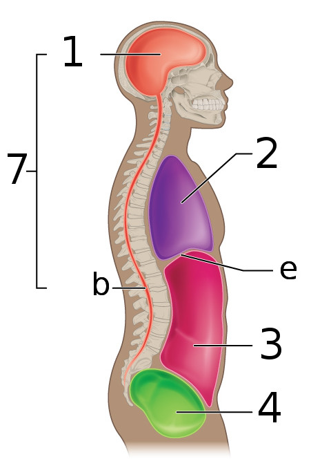
The dorsal body cavity lies towards the back of the body and is divided into two continuous parts. The cranial cavity is the space inside the skull; it contains the brain and is protected by bone on all sides. The vertebral (spinal) cavity runs inside the vertebral column and contains the spinal cord, the bundle of nervous tissue that carries signals between the brain and the body. The two cavities connect where the spinal cord leaves the skull, so they are described as one continuous dorsal cavity rather than two sealed boxes. Because the wall of this cavity is bone, bleeding or swelling inside it can press on nervous tissue, which is why a head injury or a spinal injury is treated as a serious emergency even when the skin is unbroken.
For example: The spinal cord passes through the foramen magnum, the large opening at the base of the skull, linking the cranial and vertebral cavities.
The ventral cavity: thoracic and abdominopelvic
The ventral body cavity lies towards the front and is much larger. It is divided by the diaphragm, a dome-shaped muscle used in breathing, into a superior thoracic cavity and an inferior abdominopelvic cavity. The thoracic cavity contains the two lungs and, in the middle, the mediastinum, a central region that holds the heart, the great vessels, the oesophagus and the trachea. The abdominopelvic cavity contains the abdominal cavity with the digestive organs and the pelvic cavity with the bladder, the internal reproductive organs and the lowest part of the digestive tract. Unlike the dorsal cavity, these spaces are not walled by bone alone: the thoracic cavity is protected by ribs and sternum, while the abdominopelvic cavity has muscular walls, which is why abdominal organs are more exposed to injury.
For example: A stab wound just below the left rib margin can enter the thoracic cavity if it passes upward through the diaphragm, injuring the spleen or lung base.
 Enlarge illustration ↗
Use the key: 1 cranial cavity; b vertebral canal; 7 dorsal cavity grouping; 2 thoracic cavity; e diaphragm; 3 abdominal cavity; 4 pelvic cavity. Trace the diaphragm between chest and abdomen. The abdominal and pelvic cavities are continuous, without a diaphragm between them.
Image source and licence
OpenStax · Source image · CC BY 3.0. Original image, reproduced without alteration.
Serous membranes and serous fluid
Organs inside the ventral cavity move constantly: the lungs expand, the heart beats, the intestines contract. To reduce friction, each of these organs is wrapped in a serous membrane, a thin double-layered sheet that secretes a small amount of slippery serous fluid. The layer attached to the organ is the visceral layer; the layer attached to the cavity wall is the parietal layer. Between them is a thin space containing the fluid, so the two layers slide over one another. Each region has its own name: the pleura wraps the lungs, the pericardium wraps the heart, and the peritoneum lines the abdominopelvic cavity. Inflammation of these membranes is painful precisely because the layers that normally glide can no longer move smoothly.
For example: In pleurisy, the inflamed pleura causes sharp pain when the lungs expand, because the two layers rub instead of sliding.
Common misconception: Serous membranes are not the same as mucous membranes. Serous membranes line closed body cavities and secrete lubricating fluid; mucous membranes line passages open to the exterior.
The three principal planes
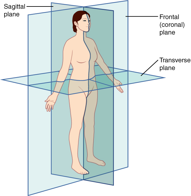
A plane is a flat surface passing through the body, and standard planes have standard names. A sagittal plane runs vertically from front to back and divides the body into right and left portions; the median (midsagittal) plane passes exactly through the midline and produces equal halves, while any other sagittal plane is parasagittal. A frontal (coronal) plane runs vertically from side to side and divides the body into anterior and posterior portions. A transverse (horizontal) plane runs horizontally and divides the body into superior and inferior portions. An oblique plane is any plane that is not parallel to the other three. Naming a plane immediately tells you what kind of view, and therefore what kind of information, a section will show.
For example: A surgeon planning an operation on the right kidney may review a frontal view to see left-to-right relationships and a transverse view to see front-to-back depth.
 Enlarge illustration ↗
Imagine each blue sheet making a cut. Sagittal separates right from left; frontal separates front from back; transverse separates upper from lower. A plane is an imaginary surface; a section is the view produced by a cut along it.
Image source and licence
Connexions · Source image · CC BY 4.0. Original image, reproduced without alteration.
From plane to section in imaging
A section is the flat surface you see when the body is cut along a plane, and every medical image is a section of some kind. A chest X-ray taken from the front is roughly a frontal projection, so it shows structures superimposed front to back. A computed tomography (CT) scan is usually a series of transverse sections, so you read it as though looking at slices from below, with the patient’s right on your left. Magnetic resonance imaging (MRI) can produce sections in any plane, which is why the radiographer states the plane in the report. Understanding planes therefore turns an unfamiliar image into a predictable map. When you look at a section, ask which plane produced it and which way the patient is facing; those two facts resolve most of the confusion.
For example: On a transverse abdominal CT, the patient’s right appears on the reader’s left, because the image is viewed from below.
Key facts to remember
- The dorsal cavity has cranial and vertebral parts and contains the brain and spinal cord.
- The ventral cavity is divided by the diaphragm into the thoracic cavity (with the mediastinum) and the abdominopelvic cavity.
- Serous membranes have a visceral layer on the organ and a parietal layer on the wall, separated by lubricating serous fluid.
- The pleura, pericardium and peritoneum are the serous membranes of the lungs, heart and abdominopelvic cavity.
- Sagittal, frontal (coronal) and transverse planes divide the body into right/left, anterior/posterior and superior/inferior portions.
Example · A paramedic suspects a collapsed lung on the left. The lung lies in the left pleural cavity, inside the thoracic part of the ventral cavity. A needle to relieve pressure is placed through the chest wall in a specific intercostal space. Understanding the cavity boundary, the pleural layers and the transverse plane lets the team name the target, avoid the structures above and below the rib, and check the result with a frontal chest image.
Read full notes
Read one topic at a time. These notes explain the mechanisms and relationships behind the key facts.
Two cavities, two kinds of protection
The dorsal and ventral cavities differ in how they protect their contents. The dorsal cavity is bony and rigid, which gives excellent protection but leaves little room for swelling: because the skull and vertebral column cannot expand, any increase in volume inside pushes on the nervous tissue. The ventral cavity is different. Its thoracic part is partly bony and partly muscular, and its abdominal part is muscular and flexible, so it can change volume as the lungs fill or the stomach stretches. The price of flexibility is greater vulnerability to direct injury. Recognising which cavity a wound or an organ lies in therefore predicts both the kind of damage likely and the kind of monitoring needed.
How serous membranes are arranged
It helps to picture each serous membrane as a deflated balloon pressed around an organ. The part of the balloon touching the organ is the visceral layer; the part against the cavity wall is the parietal layer; and the thin film of fluid between them lets the two slide. In the thorax, the pleura forms a separate sac around each lung, and the pericardium forms a sac around the heart. In the abdomen, the peritoneum is more complex because it folds around the intestines and forms sheets that suspend them. This arrangement explains several clinical signs: pain from a parietal layer is sharp and localised because that layer is supplied by somatic nerves, while pain from a visceral layer is often dull and poorly localised. Knowing which layer is irritated helps interpret what a patient describes.
Planes as a coordinate system
The three principal planes act like a coordinate system for the body. The sagittal plane controls left and right, the frontal plane controls front and back, and the transverse plane controls up and down. Any point can be located by combining positions along these three directions, which is exactly what a radiographer does when describing a lesion. The planes also standardise sections: two clinicians in different hospitals can describe the same slice in the same way, which makes comparison of images and reports possible. Learning the planes is therefore not a memory exercise but the acquisition of a shared grid that underlies every cross-sectional image you will ever see.
Why cavities are named in clinical notes
Naming a cavity quickly communicates both location and risk. “The wound entered the pleural cavity” tells a colleague that the lung may be injured and that breathing must be monitored. “The mass is retroperitoneal” tells a surgeon that the approach differs from one for an intraperitoneal structure. “The bleeding is intracranial” signals a rigid cavity and the possibility of raised pressure. Cavity names are therefore shorthand for a set of anatomical relationships and physiological dangers. When you learn a cavity, learn its boundaries and its contents, not only its name, because the boundaries determine what can enter and what can escape, and the contents determine what may be damaged.
Choosing the right plane for the question
Different clinical questions call for different planes. To see whether a structure crosses the midline, use a frontal view. To see its depth from the skin, use a sagittal or transverse view. To compare left and right at the same level, use a transverse section. To see how far something extends vertically, use a frontal or sagittal section. Trained readers rarely think consciously in these terms, but the habit is easy to build: before looking at an image, say what question you are asking, then check that the plane shown can answer it. This simple step prevents the common error of concluding that a structure is absent when it is simply outside the plane of the slice.
Explore the diagram
Start with the whole view, then select a numbered structure. Compare the additional illustrations to connect what a structure looks like with what it does.
The main body compartments
Side and front cutaways show the spaces that house the organs. Compare their boundaries and contents, one region at a time.
More controls
01 / 7 Cranial cavity
The cranial cavity is the space inside the skull. It protects the brain. The purple region in the side view continues into the vertebral canal.
Cranial cavity
The space enclosed by skull bones contains the brain and its coverings.
Three ways to section the body
The translucent sheets represent planes, not anatomical structures.
More controls
01 / 3 Median sagittal plane
A plane is an imaginary flat surface. The median sagittal plane passes through the midline and divides the body into equal right and left halves. Other sagittal planes run parallel to it.
Median sagittal plane
A vertical plane through the midline separates equal right and left halves.
Illustration credits
- Goran tek-en · Male front 3d-shaded human illustration.svg · CC BY-SA 4.0.

Adapted for this lesson: labels removed, artwork cropped or recoloured, and interactive teaching overlays added. Adapted artwork retains its source licence.
Connect the illustrations to the key ideas
DORSAL CAVITY · cranial and vertebral parts
Bone surrounds the brain and spinal cord, so swelling has little room and can compress nervous tissue.
VENTRAL CAVITY · thoracic (with mediastinum) and abdominopelvic
The diaphragm divides the two parts; serous membranes lubricate the moving organs.
PLANES AND SECTIONS · sagittal, frontal, transverse
Each plane cuts the body in a standard direction and produces a predictable kind of section.
Core Concepts Defined
- Body cavity
- A space that contains or surrounds internal structures.
- Dorsal cavity
- The back cavity, containing the cranial and vertebral cavities.
- Cranial cavity
- The space inside the skull that contains the brain.
- Vertebral (spinal) cavity
- The space inside the vertebral column that contains the spinal cord.
- Ventral cavity
- The front cavity, divided into thoracic and abdominopelvic parts.
- Thoracic cavity
- The chest cavity, containing the lungs and the mediastinum.
- Mediastinum
- The central region between the lungs, containing the heart, great vessels, oesophagus and trachea.
- Abdominopelvic cavity
- The lower ventral cavity, containing the abdominal and pelvic cavities.
- Diaphragm
- The dome-shaped muscle that separates the thoracic and abdominopelvic cavities and drives breathing.
- Serous membrane
- A thin double layer that lines a closed cavity and secretes lubricating serous fluid.
- Parietal layer
- The part of a serous membrane attached to the cavity wall.
- Visceral layer
- The part of a serous membrane attached to the organ surface.
- Serous fluid
- The slippery fluid between the two layers of a serous membrane that reduces friction.
- Pleura
- The serous membrane surrounding each lung.
- Pericardium
- The serous membrane surrounding the heart.
- Peritoneum
- The serous membrane lining the abdominopelvic cavity.
- Sagittal plane
- A vertical front-to-back plane dividing the body into right and left portions.
- Median (midsagittal) plane
- The sagittal plane exactly through the midline, producing equal halves.
- Frontal (coronal) plane
- A vertical side-to-side plane dividing the body into anterior and posterior portions.
- Transverse plane
- A horizontal plane dividing the body into superior and inferior portions.
- Oblique plane
- Any plane not parallel to the sagittal, frontal or transverse planes.
- Section
- The flat surface seen when the body is cut along a plane.
Go deeper · worked example
Reading a transverse abdominal scan
- Identify the plane. The scan is labelled transverse (axial), so it is a horizontal slice dividing the body into superior and inferior parts.
- Orient the image. Transverse images are conventionally viewed from below, so the patient’s right side appears on the left of the picture and the anterior surface is towards the top.
- Name the cavity and its contents. This slice lies in the abdominal part of the abdominopelvic cavity, lined by peritoneum, containing loops of intestine and, on this slice, the liver on the right.
- Relate a finding to the cavity. A fluid collection next to the liver lies within the peritoneal cavity, while one behind the peritoneum is described as retroperitoneal, an important distinction for planning drainage.
- Confirm with another plane if needed. A frontal or sagittal view can show whether the finding extends upward towards the diaphragm or downward towards the pelvis.
Explain it yourself · Explain why a transverse section and a frontal section of the same abdomen show different information.
Reveal a model explanation
Each plane cuts the body in a different direction, so each reveals different relationships. A transverse slice shows left-to-right and front-to-back relationships at one height, which is useful for depth and for locating a structure relative to the cavity wall. A frontal slice shows left-to-right and superior-to-inferior relationships at one depth, which is useful for seeing how far a structure extends upward or downward. Reading both builds a three-dimensional picture from two-dimensional images.
Check
Answer three questions from this section — one easy, one medium and one hard. If you miss one, you get a fresh set with explanations for the last attempt.
Practise this sectionOpen all questions and filter by difficulty or Bloom levelReferences and review status
OpenStax, Anatomy and Physiology 2e, 1.6 Anatomical Terminology (body cavities, serous membranes and planes); Tortora & Derrickson, Principles of Anatomy and Physiology, 12e, ch. 1. Author review only; medical and French-language expert review pending.
Optional further reading · English · internet required:
- Anatomy and physiology: an overview
- Structural organisation of the body
- Homeostasis and feedback
- Anatomical terminology
Original teaching schematics. English and French wording is author-reviewed for study; anatomy and French-language expert review is pending. Checks are practice only, not a graded examination.
Practice progress is saved on this device for the current learner.