SECTION 1.7
Abdominal regions and quadrants
Map the four quadrants and nine regions of the abdomen and use them to describe the site of an organ or pain.
Learn
The abdomen contains many organs packed closely together, and most of them cannot be seen or felt directly. To describe where something is, clinicians divide the abdomen with imaginary lines. The simplest division uses two lines crossing at the navel to create four quadrants: right upper, left upper, right lower and left lower. A more detailed division uses four lines to create nine regions, each with its own name. These grids do not replace anatomy, but they give a shared language for recording pain, describing a wound or pointing a colleague to the right place quickly. Learning which organs lie in which quadrant also lets you predict what a pain in a particular area may mean, and which structures you must consider before you examine the patient.
Key Concepts & Topics
Why the abdomen is divided
The abdominal cavity is the largest cavity of the body and holds organs of several systems: digestion, urinary excretion and, in the pelvis, reproduction. Unlike the chest, the abdomen has no complete bony cage; its walls are muscular, and the position of organs shifts with breathing, posture, meals and body size. A single named point is therefore less useful than a region. Dividing the abdomen into standard areas gives a stable reference that does not depend on the individual, so a note written in one hospital means the same thing in another. The divisions also help with examination: each area suggests which organs to consider and which way to move the examining hands, and they guide where a stethoscope, ultrasound probe or surgical incision is placed.
For example: The liver sits under the right rib cage and moves several centimetres with breathing, which is why its edge is described relative to the rib margin rather than to a fixed point on the skin.
The four quadrants
The four-quadrant system uses two lines that cross at the umbilicus. One runs vertically along the midline, the median plane; the other runs horizontally through the navel, the transumbilical plane. They create the right upper quadrant (RUQ), left upper quadrant (LUQ), right lower quadrant (RLQ) and left lower quadrant (LLQ). Remember that right and left are the patient’s sides. The RUQ contains most of the liver, the gallbladder and part of the duodenum; the LUQ contains the stomach, the spleen and part of the pancreas; the RLQ contains the caecum, the appendix and the lower part of the ascending colon; the LLQ contains the lower descending and sigmoid colon. The four-quadrant system is quick and widely used, but because it is coarse, it is paired with the nine-region system when more precision is needed.
For example: Appendicitis classically causes pain in the right lower quadrant, although it often begins as vague pain around the umbilicus before localising.
Common misconception: The quadrants are the patient’s right and left, not the observer’s. Facing the patient, their right upper quadrant is on your left.
The nine regions
For more precise description, the abdomen is divided into nine regions by four lines: two vertical and two horizontal. The vertical lines are the right and left midclavicular lines, drawn downward from the midpoint of each clavicle. The upper horizontal line is the subcostal line, drawn along the lower edge of the rib cage; the lower horizontal line is the intertubercular line, drawn between the two hip prominences (the iliac tubercles). The resulting grid has three rows and three columns. The top row is the right hypochondriac, epigastric and left hypochondriac regions. The middle row is the right lumbar, umbilical and left lumbar regions. The bottom row is the right iliac (inguinal), hypogastric (pubic) and left iliac regions. Each name describes a position, so the epigastric region is above the stomach area and the hypogastric region is below it.
For example: The epigastric region lies in the midline just below the sternum. Pain there commonly prompts questions about the stomach, the lower oesophagus, the pancreas and the liver.
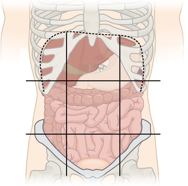 Enlarge illustration ↗
Use the navel to orient yourself. The central region is umbilical, with the epigastric region above and the hypogastric region below. The patient’s right is on your left in this anterior view.
Image source and licence
OpenStax · Source image · CC BY 3.0. Original image, reproduced without alteration.
Which organs lie where
Knowing the contents of each area turns the grid into a reasoning tool. The right hypochondriac region contains the liver and gallbladder; the epigastric region contains the stomach, the upper duodenum and part of the pancreas; the left hypochondriac region contains the spleen, the tail of the pancreas and the splenic flexure of the colon. The right lumbar region contains the ascending colon and the right kidney; the umbilical region contains loops of small intestine and parts of the transverse colon; the left lumbar region contains the descending colon and the left kidney. The right iliac region contains the caecum and appendix; the hypogastric region contains the bladder, the lower colon and, in women, the uterus; the left iliac region contains the sigmoid colon. Many organs cross the lines, so the grid narrows the possibilities rather than proving a single diagnosis.
For example: The kidneys lie high and against the posterior wall, so kidney pain is often felt in the lumbar regions and may radiate to the back or groin.
Using the grid in practice
In practice the grid structures the whole assessment. The history records where the pain started, where it is now and where it radiates; the examination moves through the regions so that no area is forgotten; and the note states the finding by region and quadrant so that another clinician can compare later. Imaging requests name the region and the suspected organ, which helps the radiographer choose the right plane and view. Surgical incisions are planned over the region containing the target organ while avoiding important vessels and nerves. The same grid also helps interpret referred pain, in which an organ produces discomfort at a distance: gallbladder pain may be felt at the right shoulder, and diaphragm irritation may be felt at the neck or shoulder tip because of shared nerve supply.
For example: A patient with right iliac fossa pain and tenderness at McBurney’s point raises suspicion of appendicitis, which is then tested with further signs and imaging.
Key facts to remember
- The four quadrants are created by the median plane and the transumbilical plane crossing at the umbilicus.
- The nine regions use two midclavicular lines, the subcostal line and the intertubercular line.
- RUQ contains most of the liver and the gallbladder; LUQ contains the stomach and spleen; RLQ contains the caecum and appendix; LLQ contains the sigmoid colon.
- Right and left always mean the patient’s sides.
- The grid guides history taking, examination, imaging requests and incision planning, and helps interpret referred pain.
Example · A patient describes pain that began around the navel and later settled in the right lower quadrant, with tenderness over the right iliac region. The quadrant tells the team to consider the appendix, the caecum and the right ureter; the nine-region detail localises the tenderness more precisely. The note records both, so that another clinician can repeat the examination and compare findings on the next shift.
Read full notes
Read one topic at a time. These notes explain the mechanisms and relationships behind the key facts.
From quadrants to regions
The two systems are not rivals; they answer questions at different scales. The four-quadrant system is fast, memorable and adequate for a first description, which is why it dominates handovers and emergency notes. The nine-region system is more precise and is used when a finding must be located carefully, such as in a wound chart or a surgical plan. A useful habit is to record the quadrant first and then the region, so that a reader who only needs the broad picture can stop reading and one who needs detail can continue. The systems share the same logic: imaginary lines create areas, and each area is associated with a set of organs. Once you know the logic, both grids are easy to reconstruct from memory.
The four quadrants and their organs
The right upper quadrant contains most of the liver, the gallbladder, the right kidney, part of the duodenum and the hepatic flexure of the colon. The left upper quadrant contains the stomach, the spleen, the left kidney, the pancreas and the splenic flexure. The right lower quadrant contains the caecum, the appendix, the lower ascending colon, the right ureter and the right ovary and uterine tube in women. The left lower quadrant contains the lower descending colon, the sigmoid colon, the left ureter and the left ovary and uterine tube. Several organs cross the midline, so the quadrant narrows the list rather than proving a diagnosis. It also sets the order of examination, because a clinician will check the structures most likely to be involved first.
Drawing the nine-region grid
To draw the grid, start with the two vertical midclavicular lines. Feel each clavicle, find its midpoint and imagine a line dropping straight down. Next add the subcostal line along the lower edge of the ribs and the intertubercular line between the hip prominences. The four lines create nine areas in three rows. The names may look unfamiliar, but each is composed of familiar roots: hypochondriac means below the cartilage of the ribs, lumbar means the loin or flank, iliac refers to the hip bone, epigastric means above the stomach and hypogastric means below it. Reading the names as descriptions of position rather than as arbitrary labels makes the grid far easier to remember.
Localising pain: local and referred
Abdominal pain does not always sit over the organ causing it. Pain from an organ’s outer covering or from the peritoneum is usually sharp and well localised, because those structures have somatic nerve supply. Pain from the organ itself is often dull, vague and felt near the midline, because visceral nerves carry fewer localising signals. Referred pain is felt at a distance from the organ, at a site that shares nerve supply with it. Gallbladder irritation may be felt at the right shoulder; diaphragm irritation may be felt at the shoulder tip or neck; kidney pain may radiate to the groin. Understanding these patterns prevents a common error: assuming that the site of the pain is always the site of the problem.
The grid in examination and documentation
A systematic examination moves through the abdomen in a fixed order so that nothing is missed. The grid supports this by giving a sequence of areas and a reason to examine each one. During documentation, findings are attached to a region so that they can be compared over time: “tenderness in the right iliac region, unchanged from yesterday” is far more informative than “still tender”. The grid also standardises communication with other services. A request for an ultrasound of the right upper quadrant tells the sonographer which region to scan and which organs to assess, while a request for the epigastric region directs attention to the stomach, pancreas and lower oesophagus. The vocabulary is small, but it carries a great deal of shared meaning.
Explore the diagram
Start with the whole view, then select a numbered structure. Compare the additional illustrations to connect what a structure looks like with what it does.
Organs of the abdomen
Anterior view of the digestive organs. Compare this with the posterior view to locate the kidneys and large vessels behind them.
More controls
01 / 6 Liver · mainly right
Liver · mainly right
The liver lies beneath the right dome of the diaphragm and extends across the midline.
Illustration credits
- Mariana Ruiz Villarreal (LadyofHats), Jmarchn · Digestive system without labels.svg · Public domain.

Adapted for this lesson: labels removed, artwork cropped or recoloured, and interactive teaching overlays added. Adapted artwork retains its source licence.
Nine abdominal regions
Read the grid from the subject’s right to left. Regions are location guides, not fixed organ boundaries.
Upper right, below the ribs; much of the liver projects here.
Connect the illustrations to the key ideas
FOUR QUADRANTS · median line + transumbilical line
Two lines crossing at the navel create RUQ, LUQ, RLQ and LLQ, using the patient’s sides.
NINE REGIONS · two midclavicular, subcostal and intertubercular lines
Four lines create three rows: hypochondriac, lumbar and iliac, with epigastric, umbilical and hypogastric in the middle.
CLINICAL LOCALISATION · name the region, then the organs
The grid guides examination, imaging requests and notes, and helps interpret referred pain.
Core Concepts Defined
- Abdominal cavity
- The space between the diaphragm and the pelvis that contains the digestive and other abdominal organs.
- Quadrant
- One of four areas made by two lines crossing at the umbilicus.
- Transumbilical plane
- The horizontal line through the umbilicus used to divide the abdomen into upper and lower halves.
- Right upper quadrant (RUQ)
- The patient’s upper right abdominal area; contains most of the liver and the gallbladder.
- Left upper quadrant (LUQ)
- The patient’s upper left abdominal area; contains the stomach and spleen.
- Right lower quadrant (RLQ)
- The patient’s lower right abdominal area; contains the caecum and appendix.
- Left lower quadrant (LLQ)
- The patient’s lower left abdominal area; contains the sigmoid colon.
- Midclavicular line
- A vertical line dropped from the midpoint of the clavicle, used to divide the nine regions.
- Subcostal line
- The horizontal line along the lower edge of the rib cage.
- Intertubercular line
- The horizontal line drawn between the two iliac tubercles, near the top of the hips.
- Right hypochondriac region
- The upper right region; contains the liver and gallbladder.
- Epigastric region
- The upper middle region, below the sternum; contains the stomach, upper duodenum and part of the pancreas.
- Left hypochondriac region
- The upper left region; contains the spleen and the tail of the pancreas.
- Right lumbar region
- The middle right region; contains the ascending colon and right kidney.
- Umbilical region
- The central region around the navel; contains small intestine and transverse colon.
- Left lumbar region
- The middle left region; contains the descending colon and left kidney.
- Right iliac (inguinal) region
- The lower right region; contains the caecum and appendix.
- Hypogastric (pubic) region
- The lower middle region; contains the bladder, lower colon and, in women, the uterus.
- Left iliac region
- The lower left region; contains the sigmoid colon.
- Referred pain
- Pain felt at a distance from the organ causing it, because of shared nerve pathways.
Go deeper · worked example
Localising right lower quadrant pain
- Place the patient in the grid. The pain is in the lower right abdomen, so it lies in the right lower quadrant, and more precisely in the right iliac region of the nine-region system.
- List the organs of that region. The caecum, the appendix, the lower ascending colon, the right ureter and, in women, the right ovary and uterine tube lie in or near the right iliac region.
- Ask about the pattern over time. Pain that begins near the umbilicus and then settles in the right iliac region is typical of appendicitis, whereas colicky pain radiating to the groin suggests a ureteric cause.
- Examine for localising signs. Tenderness at McBurney’s point, guarding and rebound tenderness all point to localised peritoneal irritation in the right iliac region.
- Record region and quadrant together. State the quadrant for a quick summary and the region for precision, then note any radiation and the timing, so the next shift can compare findings.
Explain it yourself · Explain why abdominal pain is often described by quadrant first and region second.
Reveal a model explanation
The quadrant gives a fast, shared summary that immediately suggests a set of organs and a level of urgency, so it is ideal for a first handover or a brief note. The nine regions then refine the location, because the quadrant is coarse and several organs cross its lines. Using both gives speed without losing precision: the quadrant frames the problem, and the region narrows it enough to guide examination and imaging.
Check
Answer three questions from this section — one easy, one medium and one hard. If you miss one, you get a fresh set with explanations for the last attempt.
Practise this sectionOpen all questions and filter by difficulty or Bloom levelReferences and review status
OpenStax, Anatomy and Physiology 2e, 1.6 Anatomical Terminology and 23.1 The Digestive System (abdominal regions); Tortora & Derrickson, Principles of Anatomy and Physiology, 12e, ch. 1. Author review only; medical and French-language expert review pending.
Optional further reading · English · internet required:
- Anatomy and physiology: an overview
- Structural organisation of the body
- Homeostasis and feedback
- Anatomical terminology
Original teaching schematics. English and French wording is author-reviewed for study; anatomy and French-language expert review is pending. Checks are practice only, not a graded examination.
Practice progress is saved on this device for the current learner.