SECTION 1.1
Anatomy, physiology and pathophysiology
Distinguish anatomy, physiology and pathophysiology, and explain why structure and function are studied together.
Learn
Anatomy, physiology and pathophysiology are three ways of asking about the same body. Anatomy asks what a part is and where it is. Physiology asks what that part does and how it does it. Pathophysiology asks what changes when the part or its control is disturbed, and what consequences follow. You can begin with something you already know: your hand. Its bones, joints and muscles are anatomy. The way those muscles shorten to curl your fingers is physiology. A swollen, painful finger after a sting is pathophysiology. Studying the three together is what lets a nurse predict, notice and explain a change in a patient.
Key Concepts & Topics
What anatomy studies
Anatomy is the study of the structure of the body and the relationships between its parts. Structure includes shape, size, position, and the materials a part is made from. Gross anatomy, also called macroscopic anatomy, studies structures large enough to see with the unaided eye, such as the heart, the femur or the stomach. Microscopic anatomy studies structures that require a microscope. Its two main branches are histology, the study of tissues, and cytology, the study of cells. Developmental anatomy follows how the body changes from fertilised egg to adult. Because anatomy is about position as well as form, it uses a shared vocabulary of directions and planes so that two people describe the same location in the same way.
For example: When you say the wrist is between the forearm and the hand, you are doing anatomy: you are naming parts and their positions.
What physiology studies
Physiology is the study of how the body works: the functions of its parts and the processes that keep them working. A function is the job a structure performs. Physiology asks how a muscle contracts, how a nerve carries a signal, how the kidneys remove waste, and how the body keeps its internal conditions stable. That last idea is called homeostasis, the maintenance of a steady internal environment around values the body’s cells can tolerate. Physiology often describes cause and effect: a stimulus produces a response, and the response changes the original stimulus. Physiology is usually studied one system at a time, but no system works alone, so physiology constantly links systems together.
For example: When you exercise, your heart beats faster. Explaining why that helps your muscles is physiology.
Common misconception: Physiology is not only chemistry or only movement. It includes electrical signalling, transport, temperature control and many other processes.
What pathophysiology studies
Pathophysiology is the study of how normal function is disturbed by disease or injury. It links a cause (for example, a blocked artery, an infection or a burn) to the changes that follow in cells, tissues, organs and the whole person. These changes are often called signs, which are findings another person can observe or measure, and symptoms, which are what the patient feels and reports. Pathophysiology explains not only that something is wrong but also why it produces the observations you see. This is the reasoning a nurse uses to anticipate deterioration, choose what to monitor and explain a patient’s experience.
For example: A blocked coronary artery reduces blood supply to heart muscle. The resulting chest pain is a symptom; changes on a heart tracing are signs. Linking the blockage to those findings is pathophysiology.
Structure and function belong together
The most useful habit in this course is to learn structure and function as one idea. The shape, size and material of a part help explain what it can do. A mature human red blood cell is a flexible disc without a nucleus. Its haemoglobin carries oxygen, while its flexibility helps it pass through narrow capillaries. In the lung, the thin walls of the alveoli and nearby capillaries shorten the distance gases must cross. This is the complementarity of structure and function: a part’s arrangement helps it perform its job. When you meet a new structure, ask what it is made of and how that arrangement helps it work.
For example: Your small intestine is a long tube whose inner wall is folded into finger-like projections. The folding greatly increases the surface available for absorbing nutrients — structure explaining function.
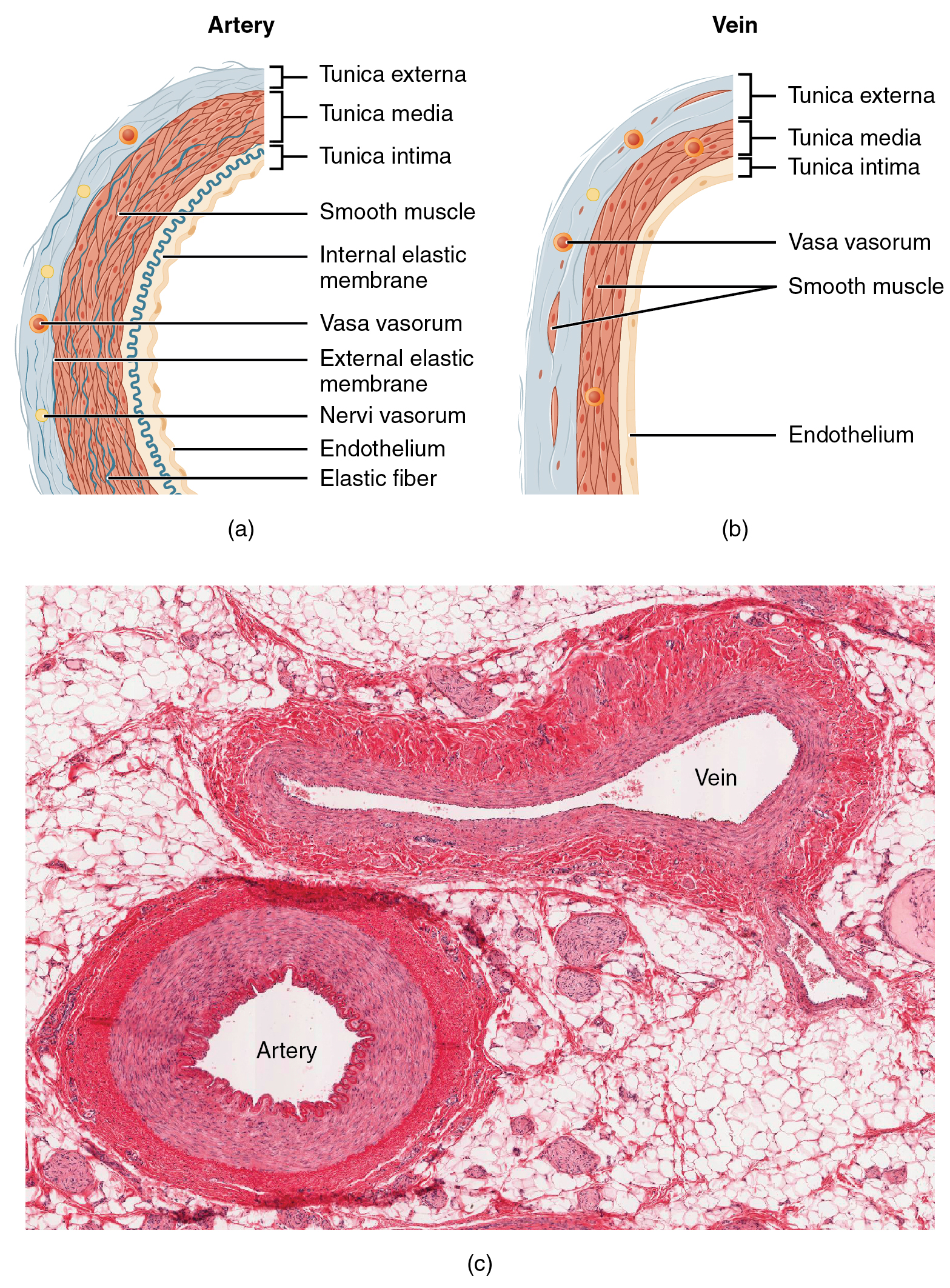 Enlarge illustration ↗
Start at the lumen—the open space carrying blood—and work outward through the layers. Compare the muscular middle layer. A vessel is a living, layered structure, not an empty rigid pipe.
Image source and licence
OpenStax College · Source image · CC BY 3.0. Original image, reproduced without alteration.
How this course uses the three
This course moves through the body system by system. For each one, you will identify the structures (anatomy), explain what they do and how they are controlled (physiology), and then see what commonly goes wrong (pathophysiology). The order matters: a mechanism is difficult to reason about before you know the parts involved. Terminology is also part of the work, because health professionals rely on precise shared words for position, direction and quantity. Learning two languages of care — the professional vocabulary and the everyday explanation a patient understands — is part of being beginner-friendly to the people you will look after.
For example: For the heart you will first locate its chambers and valves, then trace how blood moves through them, then explain what a narrowed valve does to that flow.
Key facts to remember
- Anatomy studies structure and position; physiology studies function; pathophysiology studies disturbed function.
- Gross anatomy is seen with the unaided eye; microscopic anatomy needs a microscope and includes histology (tissues) and cytology (cells).
- The complementarity of structure and function means what a part can do depends on how it is built.
- Homeostasis is the maintenance of a steady internal environment; physiology explains how it is kept.
- Signs are observed or measured by another person; symptoms are what the patient feels and reports.
Example · A patient arrives short of breath after a wasp sting. Anatomy: the airway from nose to lung. Physiology: the movement of air and the transfer of oxygen into blood. Pathophysiology: swelling narrows the airway, so less air moves and oxygen in the blood begins to fall. The same three questions organise the whole assessment.
Read full notes
Read one topic at a time. These notes explain the mechanisms and relationships behind the key facts.
Two questions, one body
Anatomy and physiology are usually taught as separate subjects, but they answer two halves of one question. Anatomy answers where and what; physiology answers how and why. Trying to explain how a muscle produces force without knowing its fibres run along its length is like explaining how a pump works without knowing it has valves. The reverse is also true: a list of bones tells you very little until you know what movement they allow and what they protect. In practice you will constantly move between the two, and the pair becomes easier with use rather than harder.
The subdivisions of anatomy
Gross anatomy can be studied region by region, such as the thorax or the abdomen, or system by system, such as the digestive system. It can also be approached by surface landmarks you can feel through the skin, which is called surface anatomy, or by the images produced by X-ray, ultrasound or scanning, called radiographic anatomy. Microscopic anatomy divides further: histology studies tissues, and cytology studies cells. Developmental anatomy follows change across the lifespan, including the rapid changes before birth. Each approach answers a different question, and clinicians choose the approach that matches the problem in front of them rather than one overall method.
From structure to function
The complementarity principle is more than a slogan. Consider three examples. Red blood cells lose their nucleus as they mature, leaving more room for haemoglobin, the iron-containing protein that binds oxygen; the cell cannot divide, but it carries oxygen well. Air sacs in the lung are one cell thick and wrapped in capillaries, so the distance gases must cross is very short. Nerve cells have long extensions that carry an electrical signal to distant targets, because a signal that must travel far needs a long conducting path. In each case the useful function follows directly from the physical arrangement, and understanding the arrangement explains both the strength and the limit of the function.
What pathophysiology adds
Pathophysiology is the bridge between a basic science and the bedside. It takes a cause — an infection, a blocked vessel, a loss of blood, a genetic change — and follows the consequences step by step through cells, tissues and organs until it reaches the observations a nurse can see. This step-by-step chain is what turns a list of facts into a prediction. If you know that a burn destroys the barrier of the skin, you can predict fluid loss, increased infection risk and pain, and therefore know what to check and what to protect. The same reasoning lets you explain to a patient not only what you are doing but why it matters, which improves both understanding and cooperation.
Talking to patients in plain language
Precision and clarity are not opposites. Professional vocabulary describes the body exactly and lets colleagues communicate without ambiguity, but a patient may not know what oedema, dyspnoea or hypertension mean. A skilled explanation uses the exact term for the record and an everyday phrase for the conversation: oedema becomes swelling, dyspnoea becomes difficulty breathing, hypertension becomes high blood pressure. This is not a simplification of the science; it is a translation of the same science into a second language. In this course each section gives both the technical term and its plain meaning, so you build the two vocabularies together from the beginning.
Explore the diagram
Start with the whole view, then select a numbered structure. Compare the additional illustrations to connect what a structure looks like with what it does.
From obstruction to tissue injury
One example of pathogenesis: obstruction reduces blood flow, which can deprive downstream tissue of oxygen.
More controls
01 / 4 Flowing blood
An open vessel delivers oxygen-bearing red cells to the downstream capillary bed. Oxygen passes from the blood towards the supplied tissue.
Flowing blood
Red cells transport oxygen along the vessel towards tissues.
Connect the illustrations to the key ideas
STRUCTURE · What is it, and where is it?
Anatomy names the parts and their positions.
FUNCTION · What does it do, and how?
Physiology explains the job and how the structure performs it.
DISTURBED FUNCTION · What changes when it is injured or diseased?
Pathophysiology links a cause to the signs and symptoms that follow.
Core Concepts Defined
- Anatomy
- The study of the structure of the body and the relationships between its parts.
- Physiology
- The study of the functions of the body and the processes that carry them out.
- Pathophysiology
- The study of how disease or injury disturbs normal function.
- Gross (macroscopic) anatomy
- The study of structures large enough to be seen without a microscope.
- Microscopic anatomy
- The study of structures that require a microscope; includes histology and cytology.
- Histology
- The study of tissues — groups of similar cells working together.
- Cytology
- The study of cells, the smallest living units of the body.
- Homeostasis
- The maintenance of a relatively stable internal environment within limits the body’s cells can tolerate.
- Complementarity of structure and function
- The principle that what a structure can do is determined by how it is built.
- Sign
- An observation or measurement made by another person, such as a rash or a raised temperature.
- Symptom
- Something the patient feels and reports, such as pain or nausea.
Go deeper · worked example
From a swollen ankle to three kinds of question
- Observe first, then classify the question. The learner sees a swollen right ankle and asks: is this a structure question, a function question or a disturbed-function question?
- Name the structures. The bones of the lower leg and foot, the joint between them, the ligaments joining bone to bone and the muscles crossing the joint are anatomy.
- Explain normal function. Ligaments hold the joint stable, muscles move it, and nerves report its position and any damage. These jobs are physiology.
- Explain what changed. A torn ligament allows abnormal movement, damaged vessels leak fluid into the tissue, and the swelling stretches pain-sensitive structures. That chain from injury to observation is pathophysiology.
- Decide what to monitor and why. Because swelling can press on vessels and nerves, the nurse checks colour, warmth, movement and sensation, then records changes over time rather than one single reading.
Explain it yourself · Explain in your own words why the same swollen ankle needs both a physiology and a pathophysiology explanation.
Reveal a model explanation
Physiology tells you what the ankle is supposed to do — bear weight, stay stable, move and report sensation — so you know what normal looks like. Pathophysiology tells you how the injury disturbs those jobs, for example a torn ligament allowing abnormal movement and leaky vessels causing swelling. Knowing both lets you predict which observations matter and what may happen next.
Check
Answer three questions from this section — one easy, one medium and one hard. If you miss one, you get a fresh set with explanations for the last attempt.
Practise this sectionOpen all questions and filter by difficulty or Bloom levelReferences and review status
OpenStax, Anatomy and Physiology 2e, 1.1 Overview of Anatomy and Physiology, 1.2 Structural Organization of the Human Body; Tortora & Derrickson, Principles of Anatomy and Physiology, 12e, ch. 1. Author review only; medical and French-language expert review pending.
Optional further reading · English · internet required:
- Anatomy and physiology: an overview
- Structural organisation of the body
- Homeostasis and feedback
- Anatomical terminology
Original teaching schematics. English and French wording is author-reviewed for study; anatomy and French-language expert review is pending. Checks are practice only, not a graded examination.
Practice progress is saved on this device for the current learner.