In one sentence Myelopathy is injury to the spinal cord caused by compression, and it is named for the region of the spine where the compression sits.
The spinal cord is a cable inside a bony canal, and compression of it is myelopathy
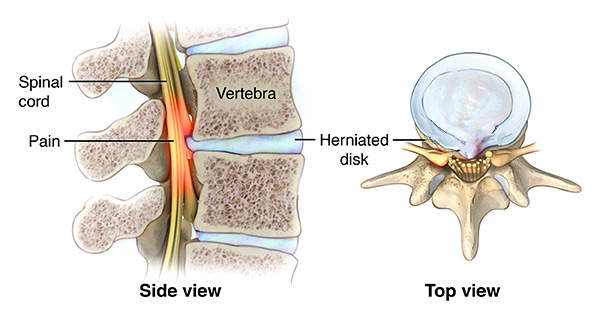
The spinal cord is a bundle of nerve fibres housed inside the vertebral canal. In adults it usually ends near vertebral L1–L2; below it, nerve roots continue as the cauda equina. When any part of that cord is squeezed or constricted, the resulting loss of function is called myelopathy. The squeezing may be sudden, as after a spinal injury, or slow, as when years of degeneration narrow the canal, and the words used to describe it join the region to the cause. Cervical myelopathy affects the cord in the neck and thoracic myelopathy affects it in the thoracic spine. Low-back compression below the cord’s endpoint affects the cauda equina or nerve roots rather than the cord itself; compression near the endpoint may affect the conus medullaris. Cervical spondylotic myelopathy is the commonest of these in older adults: spondylosis means the wear-and-tear degeneration of the discs and joints of the spine, and it narrows the neck canal around the cord. The region matters because the cord is organised from top to bottom, so a compression in the neck produces its signs in the neck, arms and hands, while a compression lower down produces them in the legs and trunk. That single rule — the level of the lesion decides the distribution of the signs — is the most useful thing to carry away from this idea.
A 68-year-old with years of neck stiffness develops clumsy hands and an unsteady walk. An MRI shows a narrowed cervical canal pressing on the cord: cervical spondylotic myelopathy.
What is myelopathy, and what does the name of each type tell you?
Show the answer
Myelopathy is injury of the spinal cord from compression or disease of the cord; each type is named for the region of the spine affected, and that region predicts where the signs will appear.
Read detailed explanations
- Tracts, reflexes and the level: why a squeezed cord gives a mixed picture
The cord is a stack of named pathways, and knowing which one is squeezed explains each sign. The corticospinal tract carries voluntary movement commands downward from the motor cortex; it crosses to the opposite side in the medulla, so a cord lesion affects the limb on the same side below the level. The spinothalamic tract carries pain and temperature upward and crosses within the cord, so it can be affected one or two segments below the level on the opposite side, producing a suspended sensory level. The dorsal columns carry vibration sense and joint position sense and cross in the medulla, so they follow the same side as the corticospinal tract. Because a degenerating neck compresses the cord from the front, through a bulging disc, and from behind, through a thickened ligament, it can catch several of these pathways at once.
The classic result is a mixed picture at a single spinal segment: lower motor neuron signs exactly at the level — weakness, muscle wasting, fasciculation and a reduced reflex, because the anterior horn cells or their roots are damaged where they leave the cord — combined with upper motor neuron signs below it, where hyperreflexia, spasticity, clonus and an extensor plantar response (Babinski sign) appear because the local reflex arcs are released from the cortex’s braking influence. Bladder control fails in two stages: first urgency and frequency, because the same release makes the detrusor overactive, and later retention with overflow. Movement is also affected by the sensory loss, because a patient who cannot feel where the foot is placed has an unsteady, wide-based gait even when strength is fair.
The examination is therefore not a hunt for one sign but a map of the level: the segment where wasting and reduced reflexes begin marks the lesion, and everything below it belongs to the upper motor neuron pattern.
- Imaging and electrical tests: what each study can and cannot show
The tests do different jobs, and choosing between them is a clinical decision. A plain radiograph shows bone: alignment, fractures, congenital narrowing and the bone spurs of spondylosis, but the spinal cord and the discs are invisible on it, which is why a normal X-ray never excludes myelopathy. Magnetic resonance imaging is the investigation of choice because it shows the discs, the ligament, the cerebrospinal fluid around the cord, the cord itself and any tumour, cyst or haematoma. It also shows signal change within the cord, a brighter area on the T2 sequence that marks oedema or established damage, and it can count how many levels are narrowed.
Myelography, done with fluoroscopy, injects iodinated contrast into the subarachnoid space around the cord and then takes images as the contrast outlines the cord and nerve roots; a block appears as a column that stops. It remains useful when a patient cannot enter a magnetic scanner — a pacemaker, a cochlear implant or a metallic foreign body in the eye — and it is combined with computed tomography for detail, but it is invasive, it can cause headache, nausea and, rarely, seizures, and it is avoided where there is infection at the puncture site or raised intracranial pressure. Electrical testing answers a different question: not where the block is, but how the nerves are conducting.
The electromyogram records the electrical activity of muscle at rest and during contraction and can show denervation, while somatosensory evoked potentials send a small electrical stimulus to a limb and record the response over the sensory cortex, tracing the signal through the whole pathway. Evoked potentials are useful for confirming a diagnosis, for monitoring the cord during decompression and for following a patient whose scans are equivocal. Decompression itself has variants: an anterior approach removes the disc and fuses that segment, while a posterior laminectomy removes the lamina and may be combined with fusion if the spine is unstable.
The nurse’s part is the same in all of them: baseline and repeated neurological observations, an airway and wound check after an anterior neck operation, and immediate reporting of a new haematoma, a falling level of consciousness or any deterioration in movement, sensation or bladder control.
- From warning to disease: how the pain system changes, and the corrected time boundary
Acute and chronic pain differ in more than duration, because the nervous system itself changes. A nociceptor is a free nerve ending that responds to mechanical, thermal or chemical injury; when tissue is damaged it releases inflammatory substances that lower the threshold of these endings, so they fire more readily. The signal travels to the dorsal horn of the spinal cord along two kinds of fibre: fast, myelinated A-delta fibres produce the sharp, well-localised first pain, and slow, unmyelinated C fibres produce the dull, aching second pain that lingers. From the dorsal horn the signal crosses and ascends in the spinothalamic tract to the thalamus and then the cortex, where it becomes the experience a person reports.
The dorsal horn is also a gate: input arriving on large touch fibres, and descending signals from the brain, can inhibit transmission there, which is the mechanism behind the relief obtained by rubbing a painful area, applying heat or cold, using transcutaneous electrical stimulation, or deliberately shifting attention. When pain signals keep arriving, the gate can be altered in the opposite direction. Central sensitisation is the change in which dorsal horn neurons become more excitable, respond to weaker input, enlarge the area they serve and fire spontaneously; wind-up is the same process measured in the laboratory, as each repeated stimulus produces a larger response. Clinically this is why pain outlasts the healed tissue, why a light touch can hurt (allodynia) and why the pain spreads beyond the original site.
Pain that begins with injury to a nerve rather than to tissue is called neuropathic, and it is characteristically burning, shooting or electric, with numbness and tingling; it responds poorly to paracetamol and non-steroidal anti-inflammatory drugs, which act on inflammation, and is treated with adjuvant drugs. The tricyclic antidepressants and the anticonvulsants work here: they dampen the abnormal firing of sensitised nerves and strengthen the descending pathways that use serotonin and norepinephrine, which is why a drug developed for depression or epilepsy also reduces pain. The duration classification describes persistence, not whether tissue damage is still present.
Chronic pain persists or recurs for more than three months and includes pain associated with ongoing disease as well as pain continuing after healing. Assess symptoms, function, mood and safety from the first presentation; the three-month classification is not a reason to postpone care. Neuropathic describes a mechanism involving a lesion or disease of the somatosensory nervous system, whereas acute and chronic describe time course: these categories can overlap.
- Caring for a patient with chronic pain: assessment, stewardship and the wider spiral
Management is a ladder, and the nurse’s work sits at every rung. Assessment comes first, because pain is what the patient says it is. A structured assessment records the site, the quality (throbbing, shooting, burning, stabbing), the severity on a scale of one to ten, the timing and pattern, what triggers it and what relieves it and, crucially, what the pain stops the patient doing: sleeping, walking, working, caring for family. Two short tools make this practical. The numerical rating scale measures severity, and a function measure such as the three-item PEG scale — pain intensity, interference with enjoyment of life, interference with general activity — is quick enough to repeat on a ward round.
The diagnostic questions map onto this exactly: when did it start, where is it, what does it feel like, how severe is it, what makes it worse, and what has helped. Imaging is used to find a treatable cause, not to measure pain, and a normal scan does not make the pain unreal. Because chronic pain syndrome carries documented consequences, the assessment is also a safety assessment: ask directly about sleep, appetite, mood and thoughts of suicide, and ask about alcohol and other drugs, because dependence, family breakdown and suicidal thoughts are among its effects. Treatment is multimodal, which means several treatments with different mechanisms at once.
Non-drug measures come first and stay: rest of the affected part in the acute phase, then heat or cold, physiotherapy and graded exercise to reduce spasticity, joint contractures and muscle wasting, occupational therapy, massage, whirlpool, acupuncture, local electrical stimulation, and the psychological methods — relaxation training, meditation, hypnosis, biofeedback, behaviour change, individual psychotherapy and group therapy. Medicines are then layered according to the type of pain: paracetamol and non-steroidal anti-inflammatory drugs for nociceptive pain; adjuvant antidepressants and anticonvulsants for neuropathic pain; opioids for severe pain, at the lowest effective dose and for the shortest necessary period, with a clear review, because these drugs carry a potential for abuse and dangerous side effects.
Opioid stewardship is a nursing responsibility: check the indication, watch for sedation, slowed breathing and constipation, ensure a laxative and an antiemetic are prescribed, teach the patient never to combine opioids with alcohol or sedatives, and know how to recognise and respond to over-sedation. Interventional options — nerve blocks, trigger point injections and epidural steroid injections — are used for specific problems such as muscle spasm or radicular pain, and they complement rather than replace the programme. Finally, remember that tolerance, physical dependence and addiction are three different things: tolerance means the same dose gives less relief, dependence means the body adapts and sudden stopping causes withdrawal, and addiction is a compulsive pattern of use despite harm. Confusing them leads nurses to under-treat pain or to stop treatment abruptly, and neither is safe.