← Common Core coursesNU 245 · Medical Pathology II
NU 245 · NursingSection 3.1 · Neurological disorders
Section 13 of 18

Neuromuscular disorders: recognition and diagnosis

Explain what a neuromuscular disorder is, sort the signs from the disease names, trace weakness back to the level of the nerve or the muscle that has failed, and say what each diagnostic test proves.

New to this course? How each section works
  1. Learn gives you one short idea at a time. Each idea starts with a single sentence, then explains it, gives an example, and asks you a small question you can answer straight away.
  2. Explore shows a diagram of the same ideas. Click the numbered parts to read what each one is.
  3. Check asks a few questions. Answer them all correctly to finish the section and unlock the next one.
  4. “Go deeper” is optional. It adds mechanism and extra detail for when you are ready — nothing you need for the checks is hidden there.
Why this matters

Neuromuscular disorders are among the few conditions in which a nurse can watch a patient lose the ability to swallow, speak or breathe within a single admission. Their early signs are ordinary — tripping, dropping objects, a droopy eyelid, tingling feet — and they are easy to dismiss until a crisis reveals them. If you can trace each sign to the level of the nerve or the muscle that has failed, you will recognise the pattern, ask for the right test, and protect the functions that fail first. This section gives you that reasoning before the named diseases arrive.

What you need to know first

You need only the everyday ideas that nerves carry messages, that muscles move the body when told to, and that a muscle shrinks when it is not used or not stimulated. The words neuron, motor neuron, atrophy and neuromuscular junction are all defined below, so no previous neurology course is assumed.

Words to know (5)
Neuron
The nerve cell, the basic unit of the nervous system, which receives, processes and sends electrical messages along its long fibre, the axon. A motor neuron carries its message from the spinal cord to a muscle fibre.
Motor neuron
The neuron whose axon leaves the central nervous system and commands a voluntary muscle; it and the muscle fibres it supplies form one motor unit. When a motor neuron dies, every muscle fibre it commanded loses its signal.
Atrophy
The wasting or shrinking of a tissue because its cells shrink or are lost; in muscle it follows the loss of nerve supply or prolonged disuse. The muscles between the thumb and index finger waste visibly after a long-standing nerve injury.
Neuromuscular junction
The microscopic relay where the end of a motor nerve releases a chemical messenger that carries the signal across to the muscle fibre. In myasthenia gravis the relay itself is attacked, so strength fades with use.
Electromyography (EMG)
A test in which a fine needle electrode records the electrical activity inside a muscle, showing whether the muscle is diseased or has lost its nerve supply. EMG can show denervation, the loss of the nerve supply, before the weakness is obvious to the patient.
One idea at a timeIdea 1 / 5

In one sentence Neuromuscular disorders begin when the neurons that command voluntary muscle become unhealthy or die, so the message never reaches the muscle and it weakens and wastes away.

A nerve signal is a chain, and any broken link weakens the muscle

A nerve signal is a relay, not a single event. A motor neuron sits in the spinal cord or the brainstem, and its long fibre, the axon, runs out to the muscle. At the end of the axon the signal crosses a microscopic relay, the neuromuscular junction, and makes the muscle fibre contract. Sensory neurons run the other way, carrying touch, pain and temperature back to the brain. Because the chain has several links, a disorder can begin at any of them — the neuron, its axon, the relay or the muscle itself — but the visible result is the same: the muscle receives no reliable command. It first becomes weak, and then, having lost its supply or its use, it shrinks, or atrophies. This is a breakdown of communication between the nervous system and the muscles, and it is the single mechanism behind every disorder in this unit. Note that the muscle is often the innocent last station: the disease may lie far away from it, in the cord or in the nerve.

For example

A patient reports tripping on kerbs and dropping cups, and the nurse notices that the muscles between the thumb and index finger look hollow. Those muscles have not been injured; they have lost the nerve signals that kept them alive and strong.

Pause and check yourself

Why does a muscle waste away when the nerve that supplies it dies?

Show the answer

Because the neuron carries the electrical command that keeps the muscle working; without that signal the muscle cannot contract normally and it shrinks.

Read detailed explanations
Localising the lesion: four levels and the signs that separate them

Localising the lesion is the step that turns a symptom into a diagnosis. Start above the muscle. A motor unit is one motor neuron plus every muscle fibre its axon branches to supply, and the number of fibres per unit varies with the precision the movement needs: a few for an eye muscle, several hundred for a thigh muscle. Damage to the neuron itself, an anterior horn cell lesion as in amyotrophic lateral sclerosis or spinal muscular atrophy, produces lower motor neuron signs: wasting, involuntary muscle twitching called fasciculation, reduced tone, and diminished or absent reflexes, with sensation preserved.

Damage to the peripheral nerve produces much the same picture in the territory of that nerve but often adds sensory loss and pain; if many nerves are affected in a glove-and-stocking pattern the cause is a polyneuropathy, while one nerve alone suggests a mononeuropathy from compression or injury. A root lesion at the spine, a radiculopathy, follows a dermatomal strip and is usually painful. Damage above the spinal cord, the upper motor neuron, gives the opposite signs: increased tone, exaggerated reflexes, stiff and slower movement, and an extensor plantar response, without early wasting. A junction disorder gives fatigable weakness with normal tone, normal reflexes and normal sensation. A muscle disorder gives weakness that is usually proximal, with normal sensation and normal or only mildly reduced reflexes.

Electromyography separates them further: in a nerve lesion the muscle at rest shows spontaneous fibrillation potentials and the number of motor units firing is reduced, whereas a myopathic muscle shows many small, short, polyphasic units. Nerve conduction studies add the conduction velocity, normally about 50 to 60 metres per second in the arm, which falls sharply when myelin is destroyed. Because amyotrophic lateral sclerosis damages both the anterior horn cell and the upper motor neuron, the same patient can show wasting and fasciculation in the hands together with brisk reflexes and spasticity in the legs, a combination no single-site disease produces.

What each test proves, and what a normal result does not exclude

Creatine kinase is an enzyme released when muscle fibres are damaged, so it is high in muscular dystrophy and in myositis and usually normal in a disorder of the nerve or of the junction; however it also rises after exercise, after an intramuscular injection and in some healthy people, so it indicates a direction rather than a diagnosis and must be read with the clinical picture. Electromyography uses a fine needle electrode to listen to the muscle at rest and during contraction. Spontaneous activity at rest, the fibrillation potentials and positive sharp waves, signals denervated or inflamed fibres, and the shape and size of the motor unit action potentials separate myopathic from neuropathic disease.

Nerve conduction studies stimulate the nerve at one point and record the response at another; the latency and the conduction velocity distinguish a demyelinating process, in which speed collapses, from an axonal one, in which amplitude falls while speed is preserved. In suspected myasthenia gravis, repetitive nerve stimulation can show a decrement in the response, and single-fibre studies can demonstrate the variable transmission directly. Magnetic resonance imaging shows structure, and gadolinium contrast shows active inflammation, but it cannot see individual nerve fibres. A lumbar puncture examines cerebrospinal fluid for cells, protein and markers such as oligoclonal bands, and it carries a risk of post-procedure headache, so it is used where central disease is suspected.

Muscle biopsy remains the tissue proof: frozen sections with immunohistochemistry reveal missing structural proteins, inflammatory infiltrates or ragged fibres. Genetic testing has moved from single-gene tests to panels and exome sequencing, and because many of these conditions are inherited in an autosomal dominant, autosomal recessive or X-linked pattern, a positive result carries implications for parents, siblings and children, so genetic counselling is part of the test, not an afterthought. Finally, sensitivity matters: a normal test early in a disease does not exclude it, and the clinical examination remains the reference standard.

The complications that decide the outcome: swallowing, breathing and the immobile patient

Bulbar muscles are the muscles of the mouth, throat and tongue, and when they weaken the patient develops dysarthria, which is slurred speech, and dysphagia, which is difficulty swallowing. Dysphagia is dangerous because food, drink or saliva can enter the airway; the result is aspiration, and aspiration leads to pneumonia, a leading cause of death in these disorders. The nurse watches for coughing or choking during meals, a wet or gurgling voice afterwards, a rising temperature and new breathlessness, keeps the patient upright during and after eating, offers thickened fluids and small mouthfuls, and involves a speech and language therapist and a dietitian. A gastrostomy tube may be needed when swallowing becomes unsafe or weight falls. Breathing fails more quietly.

Weak diaphragm and intercostal muscles reduce the volume the patient can move, so the vital capacity falls, the cough becomes too weak to clear secretions, and carbon dioxide accumulates, producing morning headache, disturbed sleep, daytime sleepiness and orthopnoea, which is breathlessness when lying flat. These are late and easily missed signs, so ventilatory capacity is measured rather than guessed, and non-invasive ventilation, secretion clearance and avoidance of sedative drugs that depress breathing are the mainstays. Immobility brings its own predictable damage: joint contractures, pressure ulcers, deep vein thrombosis, constipation and loss of bone density.

Multidisciplinary care therefore runs alongside diagnosis from the beginning, with physiotherapy to preserve range and strength, occupational therapy to adapt the home, attention to pain, cramps, fatigue and mood, and early discussion of what the patient wants if the disease progresses. Some commonly used medicines, including certain antibiotics and anaesthetic agents, can worsen transmission at the neuromuscular junction, so every new prescription is checked against the diagnosis.
Explore the diagram

Look at the whole drawing first. Then select one numbered part and connect it to the idea you just read.

From motor neuron to muscle: where the fault can lie

Follow one command for movement from the spinal cord to the muscle fibre. A motor neuron sends the signal along its axon, the axon crosses to the muscle at the neuromuscular junction, and the muscle fibre contracts. Sensory fibres run the same route in reverse, carrying touch, pain and temperature back to the cord. A disorder can begin at any link of this chain, and the link that fails decides which signs appear and which disease is named.

New to diagrams? Read this first
  1. Look at the whole drawing once without clicking anything. Ask yourself only: what kind of picture is this (a comparison, a route, a sequence, a table)?
  2. Press Play, or use the arrows, to move through the numbered parts in order. The drawing stays still; only the highlight moves.
  3. Click a number, or its name in the list below, to read what that part is and why it matters. Use “Hide labels” when you want to test your memory.
More controls
100%

01 / 5Anterior horn cell: the lower motor neuron in the cord

From motor neuron to muscle: where the fault can lieFollow one command for movement from the spinal cord to the muscle fibre. A motor neuron sends the signal along its axon, the axon crosses to the muscle at the neuromuscular junction, and the muscle fibre contracts. Sensory fibres run the same route in reverse, carrying touch, pain and temperature back to the cord. A disorder can begin at any link of this chain, and the link that fails decides which signs appear and which disease is named.One signal, four possible points of failureAnterior horn neuronlower motor neuronPeripheral nerveaxon and myelin sheathNeuromuscular junctionnerve meets muscleMuscle fibrethe contracting tissueSensory fibres run alongsidetouch, pain, temperatureWhere each disorder acts — and one mimic to excludeAnterior horn cell — ALS and spinal muscular atrophy: the lower motor neuron in the cord degenerates.Peripheral nerve — Charcot-Marie-Tooth disease and peripheral neuropathy: axon or myelin is damaged.Neuromuscular junction — myasthenia gravis: transmission across the relay fails.Muscle — muscular dystrophy, myopathy and myositis: the fibre itself is diseased.Central nervous system — multiple sclerosis: a demyelinating disease that can mimic these patterns.Recognition is not namingDroopy eyelids, double vision, numbness and cramps are consequences,not diagnoses: they say which fibres are involved, not which disease.

If the drawing is wider than this panel, scroll it sideways. Labels stay readable; the numbered explanations below show the same ideas.

Anterior horn cell: the lower motor neuron in the cord

The anterior horn cell is the lower motor neuron: its cell body lies inside the spinal cord or brainstem, and its axon leaves the cord to end on muscle fibres; the neuron and the fibres it supplies form one motor unit. When it degenerates, those fibres lose their nerve supply, become denervated and waste, while sensation stays normal because the sensory pathway is a different set of cells. Amyotrophic lateral sclerosis and spinal muscular atrophy both begin here.

Supporting illustrations

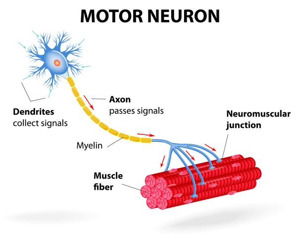
Diagram of a motor neuron: dendrites collecting signals, an axon carrying impulses, the myelin sheath, and the neuromuscular junction where the nerve meets a muscle fibre.

Figure from the supplied lecture notes: the motor neuron from dendrites to neuromuscular junction. Damage anywhere along this path — cell body, axon or junction — produces weakness, which is why these disorders are grouped by the level affected.

Original figure labels are retained. Enlarge to inspect them; the explanation is available in the selected language.

Schematic, not anatomical: the chain is drawn as four parallel stations rather than as one continuous nerve, and the sensory pathway is shown as a single branch. The anterior horn cell shown here is the lower motor neuron, whose cell body lies inside the spinal cord; the real nervous system also includes upper motor neurons, which lie above the spinal cord and are omitted here because this plate shows the nerve-to-muscle relay only.

Key points to remember

  • Neuromuscular disorders are failures of the nerve-to-muscle chain: neurons become unhealthy or die, the message fails, and the muscle weakens and atrophies.
  • Motor fibres give weakness and twitching; sensory fibres give numbness and tingling; bulbar and automatic fibres threaten swallowing and breathing.
  • Signs and disease names are mixed together, so sort them: signs are observations, diseases have a level — motor neuron, peripheral nerve, junction or muscle.
  • Diagnosis localises before it names: muscle enzymes, EMG and nerve conduction studies, MRI, lumbar puncture, muscle biopsy and genetic testing each answer one question.
  • There is no routine cure, so nursing care protects what fails first: airway and swallow, breathing, mobility, skin and quality of life.
Go deeper

Worked example

Unpacking a referral for weakness, term by term. A clinic note reads: “Six months of progressive weakness of the right hand, wasting of the first web space with visible muscle twitching, no numbness, brisk reflexes in the legs. EMG shows widespread denervation with normal sensory conduction.” Do not memorise the sentence; unpack it with the chain you have just learned.

  1. Ask whether this is nerve or muscle. Weakness with visible wasting and twitching is the signature of a lower motor neuron problem — a fault in the motor neuron in the spinal cord, whose loss causes wasting and lost reflexes — because a muscle that has lost its nerve supply wastes and fires spontaneously. In a primary muscle disease the enzyme creatine kinase is usually high and twitching is not typical, so the opening description already points away from the muscle.
  2. Ask whether sensation is involved. The note says there is no numbness, which means the sensory fibres are spared. That is important because a peripheral neuropathy usually damages sensation as well as strength, so preserved sensation pushes the lesion upward, towards the anterior horn cell in the spinal cord.
  3. Ask where the lesion is. The wasting and twitching are in the hand, so the lower motor neurons supplying the hand are failing. Yet the leg reflexes are brisk, which is an upper motor neuron sign: the pathway from the brain is also involved, and when it is cut the reflexes become stiff and brisk. One disease affecting both the lower and the upper motor neuron in the same patient is the classic pattern of amyotrophic lateral sclerosis.
  4. Ask what the tests prove. EMG denervation confirms that muscles have lost their nerve supply and tells you the process is active and widespread, not a single trapped nerve. Normal sensory conduction confirms that the sensory fibres still work, which supports a motor neuron disease rather than a neuropathy. Other tests — MRI, blood tests and genetic testing — are then used to exclude the mimics.
  5. Ask what changes for the patient. There is no routine cure, so the plan is built around function: protecting swallowing and breathing before they fail, preventing contractures, pressure injury and falls, and supporting communication, nutrition and the family. The diagnosis changes the monitoring, not just the label.

Every weakness referral can be unpacked in the same order: nerve or muscle, sensation spared or not, where in the chain, what the tests prove, and what must now be protected. Notice that the words on the page gave you the weakness and the twitching; the rest came from knowing the chain and asking each question in turn.

Common misconception

Many beginners assume that weakness always means the muscle is the problem. Muscle is the final common pathway: it contracts when told to, but the order comes from a motor neuron, travels along a peripheral nerve and crosses the neuromuscular junction. The fault may therefore lie far above the muscle, in the spinal cord or in the nerve, while the muscle is only the innocent victim that wastes for lack of a signal. This is why a patient can have severe weakness with completely healthy muscle tissue, and why sensation, reflexes and the pattern of wasting matter as much as how strong the muscle tests on examination.

Check your reasoning

A patient describes eyelids that droop and double vision that are worst in the evening, together with arm weakness that improves after a rest. Sensation and reflexes are normal. Using the chain of nerve-to-muscle transmission, explain where the fault most likely lies and why fatigue is the key clue.

Reveal an answer

The pattern points to the neuromuscular junction rather than to the nerve or the muscle. Sensation is normal, so the sensory pathway is intact, and reflexes are normal, which is unusual in nerve disease. The weakness worsens with repeated use and improves with rest, which is called fatigable weakness: the nerve releases its chemical messenger, but the relay cannot keep responding, so transmission fades as the muscle is used. Droopy eyelids and double vision are common because the small muscles of the eyelids and the eye are especially sensitive to this failure. Myasthenia gravis is the disorder that classically acts here, and it is confirmed by antibody tests and by repetitive nerve stimulation rather than by muscle biopsy, because the muscle tissue itself is healthy.

Where this comes from

  • MUSCULOSKELETAL SYSTEM PATHOLOGIES lecture notes (supplied), Level 200 nursing · Lines 849–876: neurological/neuromuscular section — the definition of neuromuscular disorders and the role of neurons in controlling voluntary muscle and carrying sensory information, the consequences of neuronal death (weakness and atrophy), the symptom list (twitching, cramps, aches and pains, muscle loss, movement and balance problems, numbness, tingling, painful sensations, droopy eyelids, double vision, trouble swallowing, trouble breathing), the list of disorders (ALS, Charcot-Marie-Tooth disease, multiple sclerosis, muscular dystrophy, myasthenia gravis, myopathy, myositis including polymyositis and dermatomyositis, peripheral neuropathy, spinal muscular atrophy), the diagnostic tests (blood enzymes, MRI, lumbar puncture, EMG, nerve conduction studies, muscle biopsy, genetic testing) and the treatment paragraph (no current cure; research into genetic therapies and new medicines; symptom treatment, delayed progression and quality of life through medication, physical therapy, occupational therapy and surgery when necessary).
  • Beyond the supplied notes: standard clinical neurology of the motor unit and its investigation · the four levels of the motor pathway and their distinguishing signs (anterior horn cell, peripheral nerve, neuromuscular junction and muscle), the motor unit and its fibre counts, lower and upper motor neuron signs, length-dependent polyneuropathy versus mononeuropathy versus radiculopathy, the myopathic versus neuropathic EMG pattern with fibrillation potentials and polyphasic motor unit action potentials, normal nerve conduction velocity of about 50–60 metres per second in the arm, creatine kinase as a muscle-damage marker with its non-specific rises, repetitive nerve stimulation and single-fibre studies in myasthenia gravis, gadolinium contrast and oligoclonal bands, muscle biopsy with immunohistochemistry, genetic inheritance patterns and counselling, bulbar and respiratory complications including aspiration, carbon dioxide retention and non-invasive ventilation, and the medicines that can worsen neuromuscular junction transmission. The supplied notes list the disorders and the tests but do not explain the mechanisms, which are uncontroversial standard neurology.
  • Correction to the supplied notes · Correction to the supplied notes: the heading “NEUROMSKLER DISORDERS” on line 849 is a spelling error and is not reproduced. The notes list multiple sclerosis among the neuromuscular disorders (line 859); multiple sclerosis is a demyelinating disease of the central nervous system that can cause weakness and sensory change but is not a disorder of the neuromuscular junction or of muscle, and it is presented here as a central nervous system mimic rather than as a neuromuscular disorder. The notes describe a blood test for “elevated enzymes” without naming them or giving units; the enzyme referred to is creatine kinase, and it is described here as a non-specific marker of muscle damage rather than a diagnostic test on its own.

Lesson progress is saved on this device for the current learner. Practice answers stay separate and are not stored.

About this course and its sources

The musculoskeletal and neurological lessons build on the supplied Level 200 nursing notes. Reproductive, respiratory, skin and nursing-response lessons use cited external references. Diagrams are original educational schematics. Sections that go beyond the supplied notes say so in their sources. Specialist clinical and French-language editorial review remain pending.