NU 245 · NursingSection 2.6 · Musculoskeletal and skin disorders
Section 8 of 18
Paget’s disease and bone tumours
Explain how Paget’s disease disturbs bone remodelling and why the affected bone is dense yet weak, describe its risk factors, symptoms, complications, tests and treatment, and distinguish benign, primary and secondary bone tumours with their usual age groups and management.
New to this course? How each section works
Learn gives you one short idea at a time. Each idea starts with a single sentence, then explains it, gives an example, and asks you a small question you can answer straight away.
Explore shows a diagram of the same ideas. Click the numbered parts to read what each one is.
Check asks a few questions. Answer them all correctly to finish the section and unlock the next one.
“Go deeper” is optional. It adds mechanism and extra detail for when you are ready — nothing you need for the checks is hidden there.
Why this matters
Paget’s disease is the clearest example in this unit of a principle that applies everywhere: a tissue that is rebuilt in the wrong pattern is weaker, not stronger, no matter how dense it looks on an X-ray. A nurse who understands that will not be reassured by a thick bone, will watch the patient with Paget’s disease for falls and fractures, and will know why a bisphosphonate is given and how it must be taken. The second half of the section matters for a different reason. Secondary, metastatic bone disease is far more common than cancer that starts in bone, and its first sign may be a fracture or a spine emergency rather than a lump. Recognising the red flags early is a nursing skill that changes whether a patient walks again.
What you need to know first
You need the idea from earlier in this unit that bone is living tissue, continually rebuilt by osteoclasts that absorb it and osteoblasts that replace it. The words remodelling, alkaline phosphatase and bisphosphonate are all defined below, so no previous oncology or pharmacology course is assumed.
Words to know (5)
Remodelling
The lifelong cycle in which osteoclasts absorb small packets of old bone and osteoblasts replace them with new bone, so the skeleton repairs itself and adapts to the loads placed on it. Because remodelling is continuous, a hormonal change can weaken the whole skeleton over months without any single injury.
Paget’s disease of bone
A chronic disorder in which remodelling becomes unbalanced and overactive, so osteoclasts remove too much bone and osteoblasts overreact, producing bone that is enlarged, deformed and weak. A femur that is larger and denser than normal on X-ray but breaks after a minor fall is typical of Paget’s disease.
Serum alkaline phosphatase
An enzyme released by osteoblasts as they build bone; its blood level rises when bone turnover is high, which makes it the standard marker of active Paget’s disease. A raised alkaline phosphatase that falls after treatment shows that the abnormal turnover has been slowed.
Bisphosphonate
A class of drug that binds to bone mineral and blocks the osteoclasts, slowing bone resorption; it is the treatment of choice for Paget’s disease. The tablet is taken fasting with water and the patient stays upright afterwards, because reflux of the drug irritates the oesophagus.
Metastatic bone disease
Cancer that began in another organ and has spread to the skeleton; it is also called secondary bone cancer and is far more common than cancer starting in bone. A patient treated for breast cancer who develops back pain and a lytic vertebra may have a metastatic deposit rather than a new bone tumour.
One idea at a timeIdea 1 / 7
In one sentence In Paget’s disease the cells that remove bone work too hard and the cells that rebuild it overreact, so the bone grows larger and denser but becomes weaker and misshapen.
Paget’s disease: the bone recycling cycle runs out of balance
Every day, normal bone is broken down and built again in a cycle called remodelling. Osteoclasts absorb small packets of old bone, and osteoblasts then replace it with new bone, so the skeleton is repaired and kept strong. In Paget’s disease this cycle shifts out of balance: the osteoclasts become more active than normal and absorb too much bone. The osteoblasts try to keep up, but they overreact and lay down an excess of new bone that is abnormally large, deformed and arranged haphazardly. The result is a bone that looks thick and dense on an X-ray but is structurally weak, so it bends, breaks and presses on the nerves and joints around it. The disease usually affects the pelvis, skull, spine and legs, and it may involve one bone or several. Its exact cause is unknown, and genetic factors, including a family history of the disease, clearly play a part.
For example
A man of 70 has an X-ray of the hip for pain. The radiologist reports that the femur is enlarged and denser than normal — and yet the bone breaks later after a minor fall, because density is not the same as strength.
Pause and check yourself
What is the basic disturbance of bone remodelling in Paget’s disease?
Show the answer
The osteoclasts become overactive and absorb too much bone, and the osteoblasts overreact by producing excess bone that is large, deformed and weak.
Read detailed explanations
Bisphosphonates: how they switch off the overactive osteoclast, and how the nurse gives them safely
Bisphosphonates are the treatment of choice in Paget’s disease because they act precisely on the cell that is overactive. The drug binds strongly to hydroxyapatite, the mineral crystal of bone, and accumulates wherever bone is being resorbed. When an osteoclast takes up the bound drug, nitrogen-containing bisphosphonates such as risedronate, alendronate, ibandronate, zoledronic acid and pamidronate inhibit farnesyl pyrophosphate synthase, an enzyme of the mevalonate pathway. That single block stops the prenylation of small signalling proteins, the osteoclast’s internal skeleton collapses, and the cell dies by apoptosis. Resorption falls, and because formation is linked to it, the osteoblasts gradually fill the defect with better-organised tissue. Two practical consequences follow for nursing.
First, absorption: an oral bisphosphonate is poorly absorbed, with a bioavailability below one per cent, and food, calcium, iron, coffee and mineral water all bind it in the gut. The tablet is therefore given on an empty stomach with plain water, and the patient must stay upright and take nothing else by mouth for at least thirty minutes, because reflux of the drug causes oesophagitis and ulceration. Second, safety: the drug lowers serum calcium, so vitamin D deficiency or unrecognised hypocalcaemia must be corrected first, and a dental review is arranged beforehand because osteonecrosis of the jaw is a rare but serious complication. An acute-phase reaction with fever, myalgia and influenza-like symptoms is common after the first intravenous dose.
Treatment is monitored by serum alkaline phosphatase, and a course is repeated when the level climbs again. Because pagetic bone is abnormally vascular, a dose given some weeks before planned surgery measurably reduces operative blood loss.
Reading the tests: alkaline phosphatase, the bone scan, the radiograph and the heart
The blood test that follows Paget’s disease is total serum alkaline phosphatase, an enzyme released by osteoblasts. It rises with high bone turnover, but the same enzyme comes from the liver and the bile ducts, so a raised value is not automatically skeletal. The nurse should look for a gamma-glutamyl transferase or a bone-specific alkaline phosphatase to separate the two sources: a normal gamma-glutamyl transferase with a high alkaline phosphatase points to bone. The level also has limits. It can be entirely normal when only one bone is involved, a pattern called monostotic disease, so a normal result never excludes the diagnosis, and it is a poor guide in a patient with liver disease.
Simple urine calcium testing is not a turnover marker: urinary calcium is usually normal in Paget’s disease, and hypercalciuria appears mainly when an active patient is immobilised. Radiographs show the shape and the internal pattern of the bone: an enlarged shaft, a thickened cortex, coarse trabeculae and mixed lytic and sclerotic areas, with the classic cotton-wool appearance in an affected skull. A radionuclide bone scan is more sensitive than a radiograph for finding every involved site, which is why it is used to map the disease before treatment.
Cardiovascular complications are explained by the same disordered tissue: pagetic bone is richly vascular, and the arteriovenous shunting inside it can raise cardiac output enough to precipitate high-output heart failure in a patient whose heart is already strained; calcification of the aortic valve has also been described. Sarcomatous transformation is the feared late event. It is rare, occurring in well under one per cent of patients, and it should be suspected when pain changes character or a known pagetic bone enlarges rapidly. The prognosis of Paget’s sarcoma is poor, so any such change is investigated urgently rather than observed.
Metastatic bone disease and the emergencies the nurse must recognise
Secondary deposits reach the skeleton through the bloodstream. The red marrow of the axial skeleton — vertebrae, ribs, pelvis, skull and the proximal ends of the long bones — is richly perfused and is the usual landing site, and the valveless vertebral venous plexus described by Batson allows tumour cells from the breast or prostate to reach the spine without passing through the lungs. Once there, deposits behave in one of two ways. Osteolytic lesions dissolve bone and are typical of myeloma and of breast, lung, thyroid and kidney cancer; osteoblastic lesions lay down abnormal new bone and are typical of prostate cancer. Both weaken the skeleton. Myeloma produces the classic punched-out lytic lesions of the skull.
A pathological fracture through a deposit may be the first sign of disease, and the femur and the vertebral column are the commonest sites. The emergency that must never be missed is metastatic spinal cord compression. The warning signs are new or worsening back pain, pain that is band-like around the chest or abdomen and worse on coughing or lying flat, weakness or heaviness in the legs, altered sensation, and difficulty passing urine or opening the bowels. Once bladder or bowel function is lost the damage is often irreversible, so suspected cord compression is treated as an emergency: the patient is kept flat, the whole spine is imaged urgently with magnetic resonance imaging, and dexamethasone is started while the results are awaited.
Treatment of bone metastases is palliative but effective: local radiotherapy for painful deposits, bone-modifying drugs such as zoledronic acid and denosumab to reduce skeletal events, analgesia by the analgesic ladder, and surgery or cement augmentation for an unstable bone. Nurses also watch for hypercalcaemia, which causes thirst, nausea, confusion and constipation, and they protect the skeleton by preventing falls, using safe moving and handling, and never twisting a patient whose spine is unstable.
Explore the diagram
Look at the whole drawing first. Then select one numbered part and connect it to the idea you just read.
Disordered remodelling and uncontrolled growth
The top band shows the balance that keeps bone strong: osteoclasts resorb a small packet of old bone and osteoblasts replace it with organised new bone. The middle band shows what happens in Paget’s disease: the osteoclasts run ahead, the osteoblasts overreact, and the bone becomes large, dense and deformed — thick enough to look strong on an X-ray, yet weak enough to bend and break. The bottom band compares a benign tumour with the two kinds of malignant bone tumour: one that begins in the bone, and the far more common one that has spread there from another organ.
New to diagrams? Read this first
Look at the whole drawing once without clicking anything. Ask yourself only: what kind of picture is this (a comparison, a route, a sequence, a table)?
Press Play, or use the arrows, to move through the numbered parts in order. The drawing stays still; only the highlight moves.
Click a number, or its name in the list below, to read what that part is and why it matters. Use “Hide labels” when you want to test your memory.
More controls
01 / 5Normal remodelling: resorption and formation in step
If the drawing is wider than this panel, scroll it sideways. Labels stay readable; the numbered explanations below show the same ideas.
Normal remodelling: resorption and formation in step
In healthy bone the two phases are coupled. Osteoclasts remove a small packet of old or damaged bone, and osteoblasts refill it with organised new bone laid along the lines of force. Because the amount removed equals the amount replaced, the skeleton keeps its shape and its strength throughout life, and a small amount of the skeleton is renewed every year. Paget’s disease is a failure of exactly this balance.
Supporting illustrations
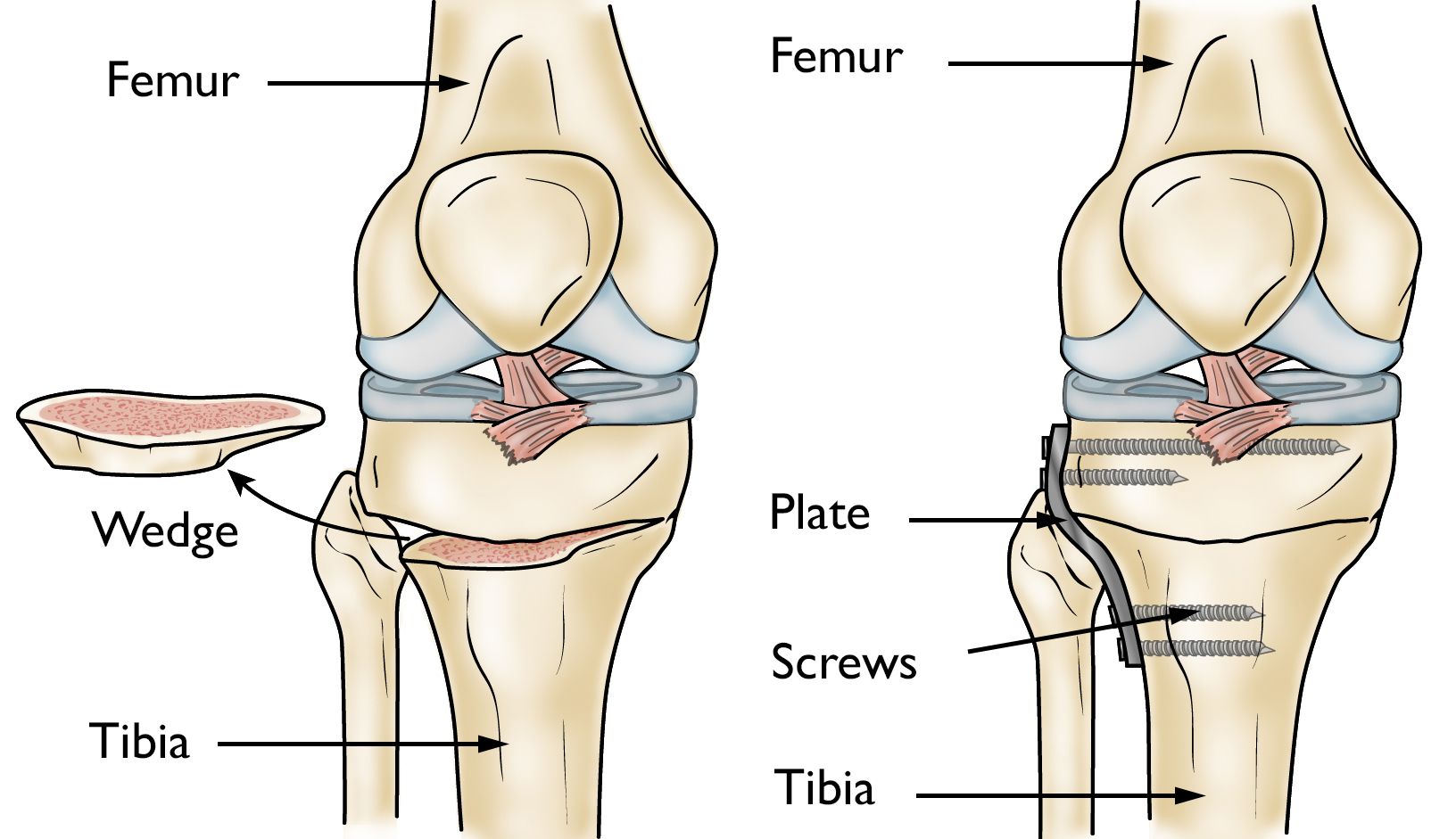
Figure from the supplied lecture notes: the tibial osteotomy used to correct the bowing that Paget’s disease can cause. The notes say the wedge is removed to restore alignment to a weight-bearing joint.
Original figure labels are retained. Enlarge to inspect them; the explanation is available in the selected language.
Schematic, not anatomical: the three bands compare processes rather than show real proportions, and the cell shapes are omitted so that the sequence stays readable. The figure also corrects two common claims: the generic cancer risk factors often listed (tobacco, obesity, alcohol, ultraviolet light) are not established for primary bone tumours, and a urine calcium test is not a marker of Paget’s bone turnover, because urinary calcium is usually normal.
Key points to remember
✓Paget’s disease is a disorder of remodelling: overactive osteoclasts and overreacting osteoblasts make bone that is larger and denser but structurally weak.
✓It is commonest after 50 and in men, is more frequent in people of northern European descent, and the symptoms depend on the bone involved — skull, spine, pelvis or legs.
✓Diagnosis uses the X-ray, serum alkaline phosphatase as the marker of bone turnover, a bone scan to map the disease and, rarely, a biopsy.
✓There is no cure; NSAIDs relieve pain, bisphosphonates block the osteoclasts and are the treatment of choice, and surgery fixes fractures, straightens deformity, replaces joints and decompresses nerves.
✓Bone tumours may be benign or malignant; malignant tumours are primary or secondary (metastatic disease from breast, lung, thyroid, kidney or prostate), and secondary disease is far more common.
Go deeper
Worked example
A 68-year-old man with night pain in the thigh: reasoning it through. A 68-year-old man has had a deep ache in the right thigh for three months. It is worse at night and he has begun taking paracetamol to sleep. His X-ray report reads: “Thickened, enlarged right femur with a coarse trabecular pattern and areas of sclerosis. No fracture.” His serum alkaline phosphatase is 340 U/L (reference 30–130), and his gamma-glutamyl transferase is normal. He is referred for a bone scan.
Start with the pain pattern. Pain that is present at rest and wakes the patient at night points to the bone itself rather than to a muscle strain, which hurts on movement and eases with rest. The three-month duration and the progressive course support a structural bone disease rather than a transient injury.
Read the X-ray description. An enlarged, thickened femur with coarse trabeculae and patches of sclerosis is the classic picture of Paget’s disease: the bone is bigger and denser than normal. The word “enlarged” is the clue, because almost no other bone disease makes a whole bone larger.
Interpret the alkaline phosphatase. Bone-building osteoblasts release alkaline phosphatase, so a value of 340 U/L fits active bone turnover. The normal gamma-glutamyl transferase matters: it makes a liver source of the enzyme unlikely, so the rise can reasonably be attributed to bone.
Map the disease with the bone scan. A bone scan shows which bones are involved and how extensive the disease is, which decides whether the patient needs treatment for one painful site or for widespread disease. It also finds silent involvement before it causes symptoms.
Set the priorities. The fracture risk comes first: the bone is weak despite its density, so falls prevention, safe handling and assistance with walking are taught now. Pain is treated with an NSAID or paracetamol plus a bisphosphonate, which blocks the osteoclasts. Before starting a bisphosphonate the nurse checks calcium and vitamin D, teaches the patient to take the tablet fasting with water and to stay upright afterwards, and asks about dental health, because osteonecrosis of the jaw is a rare complication of these drugs.
The reasoning runs in a fixed order: pain pattern, then the words of the X-ray report, then the turnover marker, then the extent of the disease, then the patient’s safety. Notice that the density of the bone never reassured anyone here — in Paget’s disease a thicker bone is a weaker bone, and the nursing work begins with preventing the fall that would break it.
Common misconception
Many beginners assume that if a bone looks dense and thick on an X-ray it must be strong, and that a “denser bone” therefore means less fracture risk. In Paget’s disease the opposite is true: the excess bone is laid down in a disorganised, haphazard pattern, so it is weaker than the normal bone it replaced and it bends or breaks under ordinary loads. Two further confusions are worth clearing up. The name is shared with unrelated conditions: Paget’s disease of bone is a disorder of remodelling, Paget’s disease of the nipple is a condition of the breast, and Paget’s sarcoma is the rare cancer that can complicate the bone disease. Finally, most people assume that cancer in bone starts in bone; in fact secondary, metastatic bone disease is far more common than primary bone cancer.
Check your reasoning
A 72-year-old man is told he has Paget’s disease of bone after an X-ray for hip pain. He asks how a bone can be “denser than normal” and still break. Using the remodelling cycle, explain it to him in plain words.
Reveal an answer
Normal bone is replaced in an orderly way: osteoclasts remove small packets of old bone and osteoblasts refill them with strong, well-organised new bone, so density and strength rise together. In Paget’s disease the osteoclasts work too fast and the osteoblasts overreact, packing in a large amount of new bone in a chaotic pattern. On an X-ray that extra tissue absorbs more radiation and looks dense, but because the fibres are not arranged along the lines of force, the bone cannot resist bending and cracking. It is like building a wall with plenty of bricks but no pattern: the wall is thick and heavy, yet it collapses under a load that a thinner, properly built wall would carry. This is why the priorities are to relieve pain, slow the turnover with a bisphosphonate and, above all, prevent the fall that would break the bone.
Where this comes from
MUSCULOSKELETAL SYSTEM PATHOLOGIES lecture notes (supplied), Level 200 nursing · Lines 519–569: Paget’s disease — the description of remodelling and the osteoclast/osteoblast imbalance, the risk factors (age over 50, male sex, origin in England, Scotland, central Europe and Greece, family history), the pelvis/skull/spine/legs distribution, the signs and symptoms by site, the diagnostic tests (X-ray, serum alkaline phosphatase, urine test, bone scan, biopsy), the complications (fracture, deformity, neurological problems, osteoarthritis, bone cancer, heart damage and hypercalcaemia), the treatment (NSAIDs, named bisphosphonates as the treatment of choice, internal fixation, osteotomy, total joint replacement, the preoperative bisphosphonate dose), and the nursing diagnoses and interventions.
MUSCULOSKELETAL SYSTEM PATHOLOGIES lecture notes (supplied), Level 200 nursing · Lines 570–595: bone tumours — the definition of an abnormal growth of cells within a bone, benign versus malignant tumours, primary versus secondary (metastatic) bone disease with breast, lung, thyroid, renal and prostate primaries, the four common primary bone cancers (multiple myeloma, osteosarcoma, Ewing’s sarcoma, chondrosarcoma) with their ages, sites and treatment, causes and risk factors, signs and symptoms, the imaging pathway (CT, MRI, PET, X-ray, bone scan) and the treatment options (amputation, radiation therapy, chemotherapy, targeted therapy, limb salvage surgery).
Beyond the supplied notes: standard bone and oncology knowledge used to explain mechanisms · bisphosphonate pharmacology (binding to hydroxyapatite, inhibition of farnesyl pyrophosphate synthase in the mevalonate pathway, apoptosis of osteoclasts), the administration rules that prevent oesophagitis, hypocalcaemia, osteonecrosis of the jaw and the acute-phase reaction, the limits of total alkaline phosphatase and the use of gamma-glutamyl transferase or bone-specific alkaline phosphatase to confirm a skeletal source, monostotic disease, the radiological terms (coarse trabeculae, mixed lytic and sclerotic areas, cotton-wool skull), high-output heart failure and aortic valve calcification in severe Paget’s disease, spread of tumour cells through the valveless vertebral venous plexus, osteolytic and osteoblastic deposits, and metastatic spinal cord compression treated as an emergency with dexamethasone and urgent MRI of the whole spine.
Corrections applied to the supplied notes · Corrections to lines 519–595: (1) the notes present a urine test showing excess calcium as evidence of rapid bone turnover, but urinary calcium is usually normal in Paget’s disease and hypercalciuria appears mainly with immobilisation, so it is not a turnover marker; (2) the notes’ risk-factor list for bone tumours (tobacco, obesity, alcohol, human papillomavirus, ultraviolet radiation) is a generic cancer list and is not established for primary bone tumours — previous high-dose radiotherapy, certain inherited cancer syndromes and Paget’s disease are the supported associations; (3) the notes call multiple myeloma the most common primary bone cancer, but it is a malignancy of marrow plasma cells rather than of bone-forming cells, and it is more accurately described as the most common primary bone malignancy in adults; (4) the notes’ incomplete phrase “It causes is unknown and genetic factors” is completed as: the cause is unknown, with a genetic contribution and a family-history risk; (5) “Paget’s sarcoma” is retained as the rare sarcomatous transformation of pagetic bone, occurring in well under one per cent of patients.
Lesson progress is saved on this device for the current learner. Practice answers stay separate and are not stored.
About this course and its sources
The musculoskeletal and neurological lessons build on the supplied Level 200 nursing notes. Reproductive, respiratory, skin and nursing-response lessons use cited external references. Diagrams are original educational schematics. Sections that go beyond the supplied notes say so in their sources. Specialist clinical and French-language editorial review remain pending.