NU 245 · NursingSection 2.4 · Musculoskeletal and skin disorders
Section 6 of 18
Sprains, strains and dislocations
Distinguish a sprain from a strain and a dislocation, describe how a force on a joint damages a ligament, a muscle or the joint surfaces, grade the severity of the injury, and apply the PRICE first-aid sequence with the nursing safety checks that go with it.
New to this course? How each section works
Learn gives you one short idea at a time. Each idea starts with a single sentence, then explains it, gives an example, and asks you a small question you can answer straight away.
Explore shows a diagram of the same ideas. Click the numbered parts to read what each one is.
Check asks a few questions. Answer them all correctly to finish the section and unlock the next one.
“Go deeper” is optional. It adds mechanism and extra detail for when you are ready — nothing you need for the checks is hidden there.
Why this matters
Sprains, strains and dislocations are the musculoskeletal injuries a nurse meets most often, in sport, at home and at work, and they are also the ones most easily dismissed as trivial. The tissue that is torn decides everything that follows: a torn ligament leaves a joint unstable and can lead to repeated dislocations and arthritis years later, while a torn muscle or tendon produces weakness that physiotherapy can rebuild. A dislocation is different again, because the displaced bone can compress a nerve or a blood vessel, and the joint must be repositioned by a doctor under sedation, never by the nurse. If you can name the injured tissue and examine the limb beyond the injury, you can give correct first aid, escalate the right patient and prevent the long-term damage that follows an injury managed badly at the outset.
What you need to know first
You need only everyday experience of a twisted ankle or a pulled muscle, and one idea already met in section 2.1: a ligament joins bone to bone, while a tendon joins muscle to bone. Every term used here is defined again below, so no previous pathology course is assumed.
Words to know (5)
Sprain
An injury in which a ligament, the fibrous band that joins one bone to another at a joint, is stretched or torn. A foot that rolls inward while running overstretches the ligaments on the outer side of the ankle.
Strain (pulled muscle)
An injury in which a muscle or its tendon is stretched too far and tears, often after overuse or a sudden effort without warming up. A sprinter feels a sudden pain at the back of the thigh: the hamstring muscle has been strained.
Dislocation
The complete separation of the two bone ends that normally meet at a joint, so that the joint surfaces no longer touch at all. After a fall on an outstretched arm, the shoulder looks deformed and the arm can no longer be moved.
Subluxation
A partial dislocation in which the joint surfaces separate only partly and then usually slip back into place on their own. A shoulder that slips partly out and back gives a feeling of giving way rather than a fixed deformity.
PRICE
The first-aid sequence started in the first 24 to 48 hours after a sprain or a strain — Protection, Rest, Ice, Compression, Elevation — to limit bleeding, swelling and further damage. A splint, an ice pack wrapped in a cloth, a compression bandage and a raised leg together make up PRICE.
One idea at a timeIdea 1 / 5
In one sentence A sprain is a stretched or torn ligament, while a strain — a pulled muscle — is a stretched or torn muscle or tendon.
A sprain injures a ligament; a strain injures a muscle or its tendon
A ligament is a tough band that runs from one bone to another across a joint and stops the joint moving further than it should. A muscle does the opposite job: it contracts to move the joint, and it transmits that pull to bone through its tendon. The same twisting force can therefore injure two very different structures, and the name of the injury tells you which one failed. A sprain is an injury to a ligament; a strain, also called a pulled muscle, is an injury to a muscle or to its tendon. The distinction is not academic. When a ligament is torn, the joint loses part of its stability and may give way under load, even though the bone is intact and the joint can look normal on an X-ray. When a muscle or tendon is torn, the joint remains stable but the patient loses strength and the movement becomes painful, especially against resistance. In everyday speech the two words are used interchangeably, which is exactly why patients arrive saying that they have “sprained a muscle”.
For example
A footballer who rolls the ankle inward tears an ankle ligament — a sprain; a runner who sprints without warming up feels a sudden pain in the calf — a strain.
Pause and check yourself
Which tissue is injured in a sprain, and which in a strain?
Show the answer
A sprain is an injury to a ligament, the band joining bone to bone, while a strain is an injury to a muscle or to its tendon.
Read detailed explanations
What happens inside the joint after a ligament tears: bleeding, inhibition and chronic instability
When a ligament ruptures, the vessels that run within it tear as well, and blood pours into the joint cavity, producing a haemarthrosis — a joint that is swollen, warm and held in a slightly flexed, comfortable position. That effusion is not only a cosmetic problem. The fluid stretches the capsule, and the swelling sends afferent signals to the spinal cord that reflexively switch off the muscle surrounding the joint, a phenomenon called arthrogenic muscle inhibition. The quadriceps, for example, may be unable to generate full force for weeks after a knee injury even though the muscle itself is untouched, and the patient reads this as instability.
At the same time, the ligament carried mechanoreceptors that reported the joint's position to the brain; when it is torn those proprioceptive signals are lost, so the joint no longer knows where it is in space and the protective reflexes fire too late. Healing itself proceeds in three overlapping phases: an inflammatory phase lasting about 72 hours, a proliferative phase in which fibroblasts lay down a disorganised collagen scar over roughly three days to six weeks, and a remodelling phase in which that scar is reorganised along the lines of stress over six weeks to as long as a year. The scar never regains the strength or the elasticity of the original ligament, and it is least tolerant of load in the first weeks. Two clinical consequences follow directly.
First, complete immobilisation protects the repair in the short term but weakens the surrounding muscle and reduces proprioception, which is why guided early movement and rehabilitation are prescribed rather than a cast for months. Second, a joint that has lost its mechanoreceptors and its ligament support keeps giving way: repeated giving-way is what defines chronic instability, and it is the pathway to further dislocation, cartilage damage and premature osteoarthritis. Nursing practice touches this at three points — controlling swelling with ice, elevation and compression during the inflammatory phase, ensuring the prescribed rehabilitation is actually attended, and warning the patient that a joint which feels normal at rest can still fail under load.
Reading the injury: what X-ray, ultrasound and MRI can and cannot show
The distinction between what a test detects and what the patient actually has is the heart of safe practice here. A plain radiograph is a picture of bone density: it shows fractures, avulsion fragments where the ligament pulled a piece of bone away, joint alignment, and, after a dislocation, that the surfaces are back in place. It does not show a ligament, so a negative film is entirely compatible with a complete tear. Stress radiographs are sometimes taken with the joint pushed in a controlled direction to reveal abnormal opening, but they need trained hands and are painful. Ultrasound, performed by a clinician, visualises ligaments, tendons and effusions dynamically — the operator can watch the tissue move — and it is quick and repeatable, though it depends heavily on the skill of the person holding the probe.
Magnetic resonance imaging is the reference method for soft tissue: it shows the ligament, the cartilage, the menisci, bone bruising and the presence of an effusion, and it is used when surgery is being considered or when the diagnosis remains unclear. The clinically important tool, however, is not an imaging machine at all. The Ottawa ankle rules are a validated decision aid: an ankle radiograph is required only if there is pain in the malleolar zone together with bone tenderness at the posterior edge or tip of either malleolus, or an inability to bear weight for four steps both immediately and in the emergency department. Applied properly, these rules reduce unnecessary radiographs without missing fractures. The nursing lessons are three.
Never tell a patient that “the X-ray was normal, so there is nothing wrong”, because that sentence contradicts everything above and discourages the rehabilitation that decides the outcome. Always record the mechanism and the examination findings, because they, not the film, establish which tissue failed. And remember that a patient with a suspected tear may be sent for further imaging after discharge, so the instructions given at that moment — who to contact, when to return, what to avoid — carry real clinical weight.
Why a dislocated shoulder is an emergency: the axillary nerve and the limits of reduction
The shoulder pays for its range of movement with a shallow socket, and that anatomy explains why it dislocates more often than any other joint — and why the structures passing close to it are at risk. The axillary nerve runs around the surgical neck of the humerus, only millimetres from the glenoid rim, and it supplies the deltoid muscle and the skin over the outer shoulder. A dislocated humeral head can stretch or compress it, so the classic findings are a patch of numbness over the deltoid and weakness when the patient tries to lift the arm. The axillary artery and the brachial plexus run nearby as well.
These structures are the reason the neurovascular examination is documented before and after reduction: a deficit that was present on arrival was caused by the dislocation, while a new deficit that appears after the manoeuvre was caused by the reduction, and the difference determines what the team does next. Reduction is performed under sedation or a short anaesthetic, both to control pain and to relax the muscles that are otherwise splinting the head in the wrong position; the sooner it is done, the less the pressure on the nerve lasts. After reduction, a radiograph confirms the position, the limb is immobilised in a sling for a period chosen by the surgeon, and the patient begins graded rehabilitation. The complication nurses must anticipate is recurrence.
Young patients, especially those under about twenty, and those whose first dislocation followed only minor trauma, have a high rate of repeat dislocation because the labrum — the fibrous rim that deepens the socket — and the capsule are torn. That combined lesion of the labrum is known as a Bankart lesion, and it may be accompanied by an indentation in the humeral head. Recurrent dislocation erodes the glenoid and damages the cartilage, so prevention is surgical or rehabilitative rather than merely expectant.
In practice, the nurse's contribution is precise and limited: immobilise in the position found, document the neurovascular state, give analgesia and keep the patient nil by mouth for the sedation, never attempt reduction, and teach the patient after discharge which activities to avoid and which warning signs — numbness, increasing pain, loss of movement — must bring them back.
Explore the diagram
Look at the whole drawing first. Then select one numbered part and connect it to the idea you just read.
Three injuries, three tissues: sprain, strain and dislocation
The same joint can be injured in three different ways, and the tissue that fails decides the signs. In a sprain the ligament that holds the bones together is stretched or torn, so the joint becomes unstable while the bone stays intact. In a strain the muscle or its tendon is torn, so the joint stays stable but strength and movement are lost. In a dislocation the two bone ends lose contact altogether, the joint is deformed and cannot move, and a nerve or vessel may be compressed. The lower band shows the five-step PRICE response that is started during the first 24 to 48 hours after a sprain or a strain.
New to diagrams? Read this first
Look at the whole drawing once without clicking anything. Ask yourself only: what kind of picture is this (a comparison, a route, a sequence, a table)?
Press Play, or use the arrows, to move through the numbered parts in order. The drawing stays still; only the highlight moves.
Click a number, or its name in the list below, to read what that part is and why it matters. Use “Hide labels” when you want to test your memory.
More controls
01 / 5Sprain: the ligament fails
If the drawing is wider than this panel, scroll it sideways. Labels stay readable; the numbered explanations below show the same ideas.
Sprain: the ligament fails
A sprain is an injury to a ligament, the fibrous band that joins one bone to another across a joint and limits how far that joint may move. The ligament may be stretched, partly torn or completely torn, and the severity depends both on the degree of the tear and on how many ligaments of that joint are damaged. Because the bone remains intact, a plain X-ray can look normal even when the joint is no longer stable, which is why a sprain is assessed by examining the joint rather than by imaging alone.
Supporting illustrations
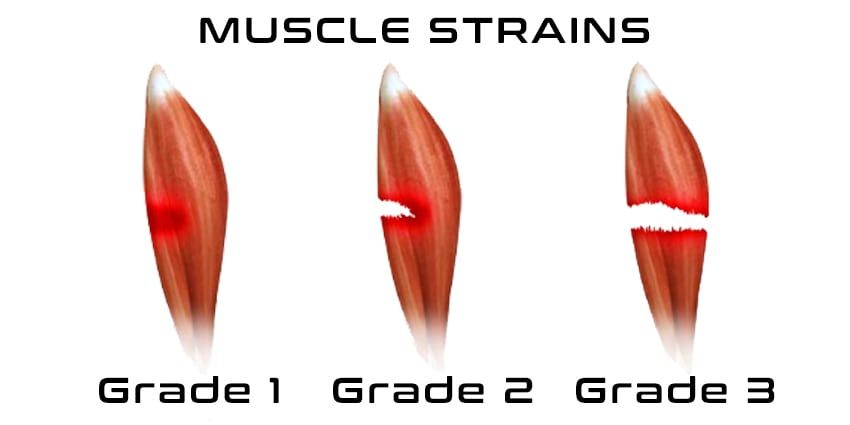
Figure from the supplied lecture notes: the three grades of muscle strain, from a stretched muscle to a torn one. A sprain is the same idea applied to a ligament.
Original figure labels are retained. Enlarge to inspect them; the explanation is available in the selected language.
Schematic, not anatomical: the three panels show the connective tissues of one joint in a simplified way, and the bone shapes are drawn as blocks rather than as real bones. PRICE is shown as a five-step chain and not as a timetable — the exact duration of each step is prescribed for the individual patient.
Key points to remember
✓A sprain injures a ligament, a strain injures a muscle or its tendon, and a dislocation separates the two bone ends at a joint completely.
✓Sprains and strains are graded as mild (stretch), moderate (partial tear) and severe (complete tear); the degree of tear and the number of ligaments involved both decide the instability.
✓The direction of the force decides the injured structure: an inward-rolling foot sprains the ankle, a blow or twist sprains the knee, a fall on an outstretched hand sprains the wrist.
✓A dislocation deforms the joint, blocks movement and can compress a nerve or vessel; the doctor reduces it under sedation, and the nurse checks pulse, sensation and movement before and after.
✓PRICE — Protection, Rest, Ice, Compression, Elevation — is started in the first 24 to 48 hours, with ice through a cloth for about 15 to 20 minutes at a time, and is followed by rehabilitation and prevention.
Go deeper
Worked example
A twisted ankle in the emergency department: from mechanism to plan. A 22-year-old student arrives on crutches after her right foot rolled inward during a game. The ankle is swollen on the outer side, bruised, and painful when she puts weight on it. She had the same injury two years ago, and she says she heard a “pop” at the moment of the twist. The doctor has ordered an X-ray.
Name the mechanism. The foot rolled inward, so the force pushed the joint in the direction opposite to the ligaments on the outer side of the ankle. Those ligaments were stretched beyond their limit: the mechanism predicts which structure is injured.
Name the tissue and the injury. The injured tissue is a ligament, and the injury is therefore a sprain, not a strain. A strain would have damaged the muscle or tendon, producing weakness and pain on contraction rather than an unstable, swollen joint.
Judge the severity. She heard a pop, cannot bear weight, and is already bruised: those findings suggest more than a simple stretch and raise the possibility of a complete tear. The degree of the tear and the number of ligaments involved together decide how unstable the joint is.
Read the X-ray correctly. The X-ray is there to exclude a fracture of the malleoli or the base of the fifth metatarsal, not to diagnose the ligament injury. If it shows no fracture, the sprain is still real; the ligament that no longer limits movement will not appear as a bone abnormality.
Plan the first hours and the follow-up. Start PRICE: protect the ankle with a splint and crutches, rest it, apply ice through a cloth for 15 to 20 minutes several times a day, compress it with a bandage and elevate it above the heart. Give analgesia as prescribed, arrange physiotherapy and teach the prevention rules, since a previous sprain is the strongest predictor of another one.
The useful order is always the same: mechanism, then tissue, then severity, then what the test can and cannot show, then the plan. Notice that this patient never needed a test to tell us that a ligament was injured; the mechanism and the examination did that, and the X-ray was requested to answer a different question.
Common misconception
Many people believe that a sprain is only a mild twist that can be walked off, and that a normal X-ray proves nothing is wrong. Both beliefs are false in the same way. A sprain is a real injury to a ligament that stabilises the joint: a complete tear can leave the joint permanently unstable, and a joint that keeps giving way is on the road to repeated dislocations and early arthritis. Because a plain X-ray shows bone rather than ligament, it cannot exclude a serious soft-tissue injury; it only excludes the fracture that might need a different treatment. Treating a sprain as trivial means skipping the protection, the rehabilitation and the strength work that decide whether the joint recovers.
Check your reasoning
A patient arrives with a shoulder that is visibly deformed after a fall, and the fingers of that hand are tingling. What will you check, what will you avoid doing, and why is this an urgent situation?
Reveal an answer
The joint is dislocated: the head of the humerus has left the glenoid cavity of the scapula, so a dislocation must be assumed until the doctor has reduced it and an X-ray has confirmed the situation. My first action is the neurovascular check beyond the injury — pulse, colour and warmth of the hand, sensation, and the ability to move the fingers — and I record it before anything is done, because a finding that is present before reduction and absent afterwards points to an injury caused by the manoeuvre. Tingling means a nerve is already being compressed, which is why the patient must be seen urgently and kept nil by mouth for a possible sedation; a numb, pale or pulseless limb would be an even more urgent finding. What I must avoid is attempting to pull the joint back into place myself: reduction is a medical procedure performed under sedation so that the muscles relax, and a nurse who tries it can damage the nerve, the vessels or the joint surfaces. I immobilise the limb in the position found, give analgesia as prescribed and keep the patient comfortable until the doctor arrives.
Where this comes from
MUSKULOSKELETAL SYSTEM PATHOLOGIES lecture notes (supplied), Level 200 nursing · Lines 355–425: the SPRAIN section (ligament definition, sprain versus strain, stretch/partial/complete severity, the ankle, knee and wrist sites, at-risk individuals, direct and indirect trauma as causes, history and physical examination, mild/moderate/severe degrees, the PRICE method with protection, rest, ice and compression, physical therapy, analgesics, anti-inflammatories and surgery, and prevention); the STRAIN section (stretched and torn muscle, causes, symptoms and first-aid ice, rest and elevation); and the DISLOCATIONS section (separation of two bones at a joint, symptoms, management by rest, ice, compression and elevation, repositioning under sedative or anaesthetic, immobilisation and surgery, and the anterior, posterior and inferior types).
Beyond the supplied notes: grading, imaging and neurovascular anatomy of joint injuries · the first-, second- and third-degree grading of ligament tears; the ligament healing phases (inflammatory, proliferative and remodelling) and their timescale; haemarthrosis and arthrogenic muscle inhibition; the Ottawa ankle rules for requesting radiographs; the limits of plain radiography with ultrasound and magnetic resonance imaging for soft tissue; the axillary nerve and the deltoid sensory patch in glenohumeral dislocation; Bankart lesions and recurrence in young patients.
Correction of the supplied notes and standard first-aid practice · Correction of the supplied notes: the notes expand PRICE with only four items (protection, rest, ice, compression) although the acronym requires a fifth, Elevation, which their own dislocation-management list includes. The notes also give the ice application as “10 minutes” and then state that ice must not be applied “for longer than 20 minutes at a time”; the authored text uses the conventional 15 to 20 minutes at a time through a cloth, with the notes’ inconsistency labelled rather than reproduced. The notes name the humerus against the glenoid when describing shoulder dislocation types; the authored text names the joint explicitly as the glenohumeral joint, formed by the head of the humerus and the glenoid cavity of the scapula.
Lesson progress is saved on this device for the current learner. Practice answers stay separate and are not stored.
About this course and its sources
The musculoskeletal and neurological lessons build on the supplied Level 200 nursing notes. Reproductive, respiratory, skin and nursing-response lessons use cited external references. Diagrams are original educational schematics. Sections that go beyond the supplied notes say so in their sources. Specialist clinical and French-language editorial review remain pending.