NU 245 · NursingSection 2.3 · Musculoskeletal and skin disorders
Section 5 of 18
Named fracture types and their management
Recognise the named fracture patterns from the force that produced them, say what each name predicts about stability and soft-tissue damage, and describe how each type is reduced, held, rehabilitated and monitored.
New to this course? How each section works
Learn gives you one short idea at a time. Each idea starts with a single sentence, then explains it, gives an example, and asks you a small question you can answer straight away.
Explore shows a diagram of the same ideas. Click the numbered parts to read what each one is.
Check asks a few questions. Answer them all correctly to finish the section and unlock the next one.
“Go deeper” is optional. It adds mechanism and extra detail for when you are ready — nothing you need for the checks is hidden there.
Why this matters
The word in front of “fracture” is not decoration. “Greenstick”, “spiral”, “comminuted”, “compression” and “stress” each tell you which force acted, how stable the break is and what is likely to happen to the patient. That name helps decide whether a cast is enough or whether the patient goes to theatre, and it warns you which complications to watch for — an open wound, a nerve injury or the limb-threatening pressure of acute compartment syndrome. Reading the name correctly is therefore a nursing safety skill, not a vocabulary exercise.
What you need to know first
You should already be comfortable with the previous section: a fracture is a break in the continuity of bone, it is classified by pattern, by open or closed skin and by displacement, and it tears the periosteum and its vessels, so the bone bleeds into the surrounding tissue. No other preparation is needed; every type below is defined where it first appears.
Words to know (5)
Greenstick fracture
An incomplete fracture of a soft child’s bone in which one side cracks while the other side only bends, like a green branch that splinters on one side. A 7-year-old falls on an outstretched arm and the X-ray shows a crack on one side of the radius only.
Comminuted fracture
A complete fracture in which the bone is broken into three or more fragments; more than four fragments is often called highly comminuted. A motorcycle crash can shatter the femoral shaft into several pieces that must be held together surgically.
Compression fracture
A fracture in which the bone is crushed along its axis, most often a vertebral body that loses height while the spine is loaded. An older woman with osteoporosis develops a wedge compression fracture of a thoracic vertebra after bending forward.
Stress fracture
A tiny crack produced by repeated load rather than by one blow, when bone is broken down faster than it can be rebuilt. A runner who doubles her weekly distance too quickly develops a stress fracture of a metatarsal in the foot.
Acute compartment syndrome
A limb-threatening rise in pressure inside a closed muscle compartment that stops the blood supply to muscle and nerve. A tight cast over a tibial fracture can raise compartment pressure until the patient loses sensation and movement in the foot.
One idea at a timeIdea 1 / 6
In one sentence A fracture name records the kind of force that broke the bone, and that force also decides how unstable and how open the injury is.
The name of a fracture is a summary of the force that caused it
Fracture names are not arbitrary labels; each one is shorthand for a mechanism. A force that bends a bone produces a line running across the shaft, or, in a soft child’s bone, a greenstick crack on one side only. A force that twists the limb produces a spiral line that wraps around the shaft like a corkscrew. A force applied along the axis compresses the bone and either jams the two ends into each other, an impacted fracture, or crushes a vertebral body. A single blow of high energy breaks the bone into several pieces, a comminuted fracture, or leaves a free segment between two fracture lines, a segmental fracture. Repeated smaller loads, arriving faster than the bone can repair itself, produce a stress fracture. Finally, a tendon or ligament pulled suddenly can tear off the piece of bone it attaches to, an avulsion fracture. Because the mechanism is written into the name, the name also predicts the soft-tissue damage, the stability and much of the treatment — which is why learning the classification pays off at the bedside.
For example
A footballer who twists a planted leg while turning gets a spiral fracture of the tibia; a recruit who runs for a week in new boots gets a stress fracture of the same bone.
Pause and check yourself
What does the name of a fracture tell you besides the shape of the break?
Show the answer
It tells you which force acted, and therefore how much soft tissue was damaged, how stable the break is and roughly how it will be treated.
Read detailed explanations
Why the direction of the force writes the fracture line
Bone is a composite material, and its behaviour under load explains every name in this section. Mineral crystals resist compression, while collagen fibres resist tension, so healthy bone is strongest when it is squeezed and weakest when it is stretched or sheared. When a limb is bent, one side of the shaft is placed under tension and the opposite side under compression; the tension side fails first, and the crack then runs across the shaft, producing the transverse line. A child’s bone contains more water and less mineral, and its periosteum is thicker, so the tension side cracks while the compression side merely bows — the greenstick pattern — and the intact sleeve of periosteum then keeps the two halves aligned.
Torsion is different: a twisting force produces shear stress distributed along the shaft, and the crack propagates as a helix around the bone, which is why a spiral fracture line is long and oblique and is often accompanied by stripped periosteum. Axial loading drives the two fragments together; if the bone is dense it may hold, giving an impacted fracture, and if the bone is a vertebra the body collapses. The energy available decides the number of fragments. A low-energy break gives two pieces, while a high-energy impact releases more energy than the bone can absorb, so the crack branches repeatedly and produces comminuted or segmental patterns with extensive soft-tissue injury. Finally, bone is viscoelastic and repairs itself continuously, so a load that is harmless once becomes damaging when repeated before remodelling can complete.
That is the mechanical basis of the stress fracture, and it is also why a bone weakened by osteoporosis, tumour or infection can break during ordinary activity — a pathological fracture, in which the force is trivial and the bone is abnormal.
Acute compartment syndrome: the complication the nurse detects first
A fracture can threaten a limb even when the bone itself is stable. Muscles, nerves and vessels are grouped into closed compartments bounded by bone and by inelastic fascia, and each compartment tolerates only a small rise in pressure. Bleeding from the broken bone, oedema from damaged muscle and external compression from a tight cast or dressing all raise the pressure inside that fixed space. As the pressure rises, the small vessels that perfuse the muscle are compressed, and because capillary perfusion depends on the difference between arterial pressure and tissue pressure, perfusion stops long before the arterial pulse disappears. Ischaemia then damages muscle and nerve, and if it continues the result is contracture, permanent weakness or amputation.
The classic warning signs are remembered as the six Ps: pain that is disproportionate to the injury and worsened by passive stretching of the muscle, a tense and swollen compartment, paraesthesia or numbness in the territory of a nerve running through it, pallor, and — late — paralysis and pulselessness. The order matters. Distal pulses are usually preserved in early compartment syndrome, so a palpable pulse is falsely reassuring, and waiting for a missing pulse means waiting until the tissue is already dying. Compartment pressure can be measured with a needle manometer; a pressure within about thirty millimetres of mercury of the diastolic pressure, or a clearly raised absolute value with typical symptoms, is an indication for urgent fasciotomy, in which the fascia is divided along the length of the compartment to release the pressure.
Nursing actions in the meantime are specific: remove or loosen any constricting bandage, splint or cast, keep the limb at the level of the heart rather than elevated above it, because elevation lowers arterial inflow without lowering compartment pressure, give the prescribed analgesia, and report the findings immediately to the treating team. The nurse who documents increasing pain and new numbness is the person who saves the limb.
Stress fractures: the remodelling lag and the people it catches
Bone responds to load by remodelling, a continuous cycle in which osteoclasts resorb a small packet of bone and osteoblasts replace it. During the resorption phase the bone is temporarily weaker, so a sudden increase in training produces a window in which the skeleton is being broken down faster than it is being rebuilt. When a muscle is fatigued it also absorbs less shock, so more of the impact is transferred to the bone beneath it; repeated cycles then concentrate stress at one point and produce a microfracture that enlarges into a stress fracture if loading continues. This mechanism explains the typical story: a new activity, a sudden rise in distance or intensity, or a change of surface and footwear, followed by pain that begins vaguely, localises to a point on the bone and eases with rest. It also explains the risk factors.
Women with absent or irregular menstrual periods have lower oestrogen, which accelerates bone resorption; low energy availability and eating disorders reduce calcium and vitamin D, the raw material of repair; flat feet, rigid high arches and worn shoes alter the way load is transmitted; and osteoporosis, previous stress fractures and a sedentary start all narrow the safety margin. Because X-rays may be normal for the first two to three weeks, a normal film does not exclude the diagnosis, and magnetic resonance imaging or a bone scan is used when clinical suspicion is strong. The principles of prevention mirror the mechanism: increase training gradually, use appropriate footwear and arch support, and maintain adequate calcium, vitamin D and energy intake.
Treatment follows the same logic by removing load — a walking boot, brace or crutches — with surgery reserved for fractures that do not heal or that occur at high-risk sites such as the anterior tibia or the femoral neck.
Explore the diagram
Look at the whole drawing first. Then select one numbered part and connect it to the idea you just read.
From the force to the fracture name to the management
The force that breaks a bone writes the shape of the fracture line, and the shape carries a name. Bending gives a transverse line, or a greenstick crack on one side of a child’s bone; twisting gives a spiral; axial compression jams the ends together or crushes a vertebra; repeated load and a sudden pull give stress and avulsion fractures. Whatever the name, management asks whether the fracture is stable, open and neurovascularly intact, then reduces, holds, rehabilitates and watches for the compartment syndrome that can threaten the limb.
New to diagrams? Read this first
Look at the whole drawing once without clicking anything. Ask yourself only: what kind of picture is this (a comparison, a route, a sequence, a table)?
Press Play, or use the arrows, to move through the numbered parts in order. The drawing stays still; only the highlight moves.
Click a number, or its name in the list below, to read what that part is and why it matters. Use “Hide labels” when you want to test your memory.
More controls
01 / 5Bending: the line that crosses the shaft
If the drawing is wider than this panel, scroll it sideways. Labels stay readable; the numbered explanations below show the same ideas.
Bending: the line that crosses the shaft
When a bone is bent, the convex side is stretched and fails before the concave side, so the crack runs across the shaft as a transverse line. In a child the mineral-poor bone and the thick periosteum let the tension side crack while the other side merely bows, producing the greenstick pattern that is most common under the age of ten. Both are usually held in a cast or splint for four to six weeks.
Supporting illustrations
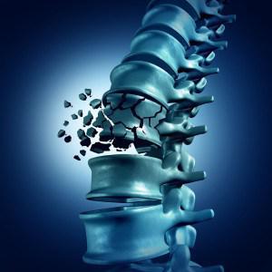
Figure from the supplied lecture notes: a compression fracture of a vertebra. Osteoporosis is its commonest cause, and the loss of height at the front of the vertebra is what produces kyphosis.
Original figure labels are retained. Enlarge to inspect them; the explanation is available in the selected language.
Schematic, not anatomical: the bone bars are simplified rectangles and each card shows only the single force that best explains that name. Real injuries combine forces — a fall from height can bend, compress and twist at once — and the same patient may have several fracture types together.
Key points to remember
✓A fracture name summarises the force that caused it, so it also predicts stability, soft-tissue damage and the likely treatment.
✓A greenstick fracture cracks one side of a soft child’s bone only, while an impacted fracture jams the broken ends together; both are usually treated conservatively with a cast or splint.
✓Transverse, oblique, spiral, comminuted and segmental fractures are complete breaks; higher energy means more fragments, more soft-tissue injury and more need for surgery.
✓In a vertebral compression fracture the shape decides the danger: a wedge is common and stable, a crush often heals without surgery, and a burst can drive fragments into the spinal cord and needs urgent care.
✓Stress fractures come from load that outruns bone repair and avulsion fractures from soft tissue pulling off a fragment; both are managed mainly by reducing load and immobilising the part.
✓Management asks whether the fracture is stable, open and neurovascularly intact, then reduces, holds, rehabilitates and monitors — above all for the rising pain of acute compartment syndrome.
Go deeper
Worked example
An older woman with sudden back pain after bending forward. A 76-year-old woman felt a sudden sharp pain in the middle of her back while reaching for a low cupboard. She has lost height over the last few years, has a stooped upper back, and the pain is worse when she stands and eases when she lies flat. A lateral X-ray shows a wedge compression fracture of T8, and a DEXA scan gives a bone density result in the osteoporotic range. She has no numbness, weakness or bladder problem.
Name the structure and its job. T8 is a thoracic vertebra of the axial skeleton. It bears load, allows the trunk to bend and twist, and, together with the vertebral canal, protects the spinal cord. Because it is a weight-bearing structure, losing height there changes the whole column, not just one bone.
Read the mechanism and the name. There was no fall and no high-energy accident; the vertebra collapsed under an ordinary movement, which points to weak bone. Osteoporosis is the commonest cause of compression fracture, and the wedge shape means the front of the vertebra failed while the back stayed intact. That pattern is usually stable, unlike a burst fracture, in which fragments are driven outward toward the canal.
Check the danger signs. The nurse asks about numbness and tingling, muscle weakness, difficulty walking and any change in bladder or bowel control, because those findings would mean nerve or cord involvement and would change the plan immediately. This patient has none, so the fracture can be managed without emergency surgery, but the level of the fracture and the alignment are recorded as a baseline.
Treat the bone as well as the fracture. The plan combines analgesia, a back brace for support, a short period of bed rest followed by graded activity and physiotherapy, and treatment of the underlying osteoporosis with medicine, calcium and vitamin D. The nurse checks that the calcium and vitamin D are actually taken, teaches safe movement — bending at the knees, not the waist — and watches for constipation from analgesia and for reduced mobility, which raises the risk of blood clots in the legs.
Plan for prevention and follow-up. If pain persists despite these measures, vertebroplasty or kyphoplasty may be offered: a needle injects quick-setting cement into the vertebra under X-ray guidance, and in kyphoplasty a small balloon first expands the collapsed space. Meanwhile the nurse reduces the risk of the next fracture by checking the home for trip hazards, reviewing medicines that cause drowsiness or dizziness, encouraging weight-bearing exercise and a diet rich in calcium and vitamin D, and arranging the follow-up that repeats the bone density measurement.
A compression fracture in an older person is not just a broken bone; it is a message that the skeleton has become fragile, and it predicts the next fracture unless the osteoporosis is treated. The nurse’s contribution is to detect nerve involvement early, to keep the patient moving safely rather than resting indefinitely, to make sure the calcium, vitamin D and bone medicine are taken, and to remove the falls and trip hazards that would turn fragile bone into the next fracture.
Common misconception
Many beginners believe that a fracture is one clean crack, that a cast is the standard treatment, and that if the limb looks straight the injury must be mild. The named patterns show how wrong that is. Some breaks are incomplete, such as the greenstick fracture, some are jammed together, such as the impacted fracture, some are shattered into several fragments, such as the comminuted or segmental fracture, and some have no visible deformity at all because they are inside a vertebra. Treatment is decided by the force, the stability of the bone ends and the state of the soft tissues, not by how dramatic the limb looks, and an apparently minor fracture can still hide an open wound, a nerve injury or the rising pressure of acute compartment syndrome.
Check your reasoning
A patient had a tibial shaft fracture placed in a cast six hours ago. He now reports severe pain that morphine has not relieved, the foot hurts when the nurse passively plantar-flexes it, and he says the top of his foot feels numb. The foot is pink and a pulse is easily felt. Using the fracture types and their management, explain what is happening and what the nurse must do first.
Reveal an answer
This is the early picture of acute compartment syndrome, not ordinary post-fracture pain. Bleeding and swelling inside a closed muscle compartment have raised the pressure until the small vessels that feed the muscle and nerve are compressed. Pain that is disproportionate and not relieved by morphine, pain on passive stretching of the muscles and new numbness are the earliest and most reliable signs; a pink foot and a palpable pulse do not exclude the diagnosis, because the arterial pulse survives long after the capillary perfusion of muscle has stopped. The nurse must act immediately rather than simply increase analgesia: remove or loosen the cast, bandage or any other constricting material, keep the limb at the level of the heart rather than raised above it, document the neurovascular findings with times, and report to the treating team at once. Compartment pressure may then be measured, and if the pressure is high enough an urgent fasciotomy is performed to divide the fascia and release the pressure before muscle and nerve die. The limb, not the X-ray, is what is at stake here, and delay is the factor that turns a recoverable injury into permanent weakness or amputation.
Where this comes from
MUSKULOSKELETAL SYSTEM PATHOLOGIES lecture notes (supplied), Level 200 nursing · Lines 151–354: named fracture types — greenstick (children under 10, causes, symptoms, X-ray/CT/MRI, cast or splint for 4–6 weeks, paracetamol or NSAID, surgery and rare complications), transverse (definition, long bones, causes, symptoms, closed reduction with local anaesthetic, sedative or general anaesthesia, internal fixation, compartment syndrome, malunion, nonunion, osteomyelitis), spiral, stress (overuse mechanism, remodelling, risk factors, prevention, walking boot or crutches, surgery), compression (osteoporosis as commonest cause, burst/crush/wedge types, symptoms including kyphosis, DEXA, kyphoplasty and vertebroplasty, complications), oblique, impacted, segmental, comminuted and avulsion fractures with their management. The notes’ spelling and labelling errors — “TRANSVERCE FRACTRE”, “MIR”, “DIGNOSIS”, “impact fracture” and “segmented fracture” — are corrected in the authored content.
Beyond the supplied notes: standard orthopaedic biomechanics and compartment syndrome · the composite behaviour of bone under tension, compression and shear; how bending, torsion and axial loading determine the fracture line; the role of the thicker paediatric periosteum; the concept of a pathological fracture; the six Ps of acute compartment syndrome and the fact that distal pulses are usually preserved early; the use of a needle manometer and the pressure threshold for fasciotomy; the nursing measures of releasing constricting material and keeping the limb at heart level; and the remodelling lag and female risk factors behind stress fractures. The supplied notes describe the fracture types and their management but do not explain the mechanics, the observation signs or the pressure threshold, which are uncontroversial standard orthopaedics. The notes also call impaction an incomplete fracture; the authored content labels this correction and explains that impaction describes jammed ends and may be a complete but stable fracture.
Lesson progress is saved on this device for the current learner. Practice answers stay separate and are not stored.
About this course and its sources
The musculoskeletal and neurological lessons build on the supplied Level 200 nursing notes. Reproductive, respiratory, skin and nursing-response lessons use cited external references. Diagrams are original educational schematics. Sections that go beyond the supplied notes say so in their sources. Specialist clinical and French-language editorial review remain pending.