← Common Core coursesNU 245 · Medical Pathology II
NU 245 · NursingSection 2.1 · Musculoskeletal and skin disorders
Section 3 of 18

Bones, joints and muscles: structure and function

Describe what the musculoskeletal system is made of and what each tissue does, name the bone cells and bone shapes, classify joints by how they move, and explain how a muscle pulls on a bone to produce movement.

New to this course? How each section works
  1. Learn gives you one short idea at a time. Each idea starts with a single sentence, then explains it, gives an example, and asks you a small question you can answer straight away.
  2. Explore shows a diagram of the same ideas. Click the numbered parts to read what each one is.
  3. Check asks a few questions. Answer them all correctly to finish the section and unlock the next one.
  4. “Go deeper” is optional. It adds mechanism and extra detail for when you are ready — nothing you need for the checks is hidden there.
Why this matters

Every disorder in this unit is a failure of one tissue you are about to learn. A fracture is a failure of bone, a sprain is a failure of ligament, arthritis is a failure of joint surfaces, and muscular dystrophy is a failure of muscle. If you can name the tissue and its normal job, you can predict what the patient will lose — support, movement, joint stability or protection of an organ — and you can read a fracture or dislocation description instead of memorising the words. This section gives you the vocabulary that the rest of the musculoskeletal and neurological units use on every page.

What you need to know first

You need only everyday experience of your own body: that a fall can break a bone, that a joint bends, and that a muscle becomes firm when it works. The names of the tissues, the bone cells and the joint types are all defined below, so no previous anatomy course is assumed.

Words to know (5)
Musculoskeletal system
The bones, cartilage, ligaments, tendons and muscles that together give the body its shape, support it, protect its organs and move it. A fractured femur disturbs both support and movement because it belongs to the same system as the muscles acting on it.
Joint
The point at which two or more bones meet, ranging from an immovable seam in the skull to a freely moving knee. The knee is a movable joint; the plates of the skull meet at immovable joints.
Ligament
A strong band of fibrous tissue that attaches one bone to another and holds a joint stable. A sprain is a stretched or torn ligament, so the joint becomes unstable rather than painful only.
Tendon
A tough cord of connective tissue that attaches a muscle to a bone so the muscle’s pull can move the skeleton. The Achilles tendon transmits the calf muscle’s pull to the heel bone.
Osteoblast and osteoclast
The bone-forming cell and the bone-removing cell; their balanced activity rebuilds bone throughout life. When osteoclasts work faster than osteoblasts, bone becomes fragile, as in osteoporosis.
One idea at a timeIdea 1 / 5

In one sentence The skeleton supports the body, lets muscles move it, protects vital organs and makes blood cells in its marrow.

One system, four jobs: support, movement, protection and blood production

Bones, cartilage, ligaments, tendons and muscles work as one system, so a problem in any one of them shows up as a problem in the others. The skeleton gives the body its shape, holds it upright against gravity and provides the levers that muscles pull on to produce movement. It also protects: the skull surrounds the brain, the vertebral column surrounds the spinal cord, and the rib cage surrounds the heart and lungs. Less obvious but equally important, some bones contain marrow that produces red blood cells, white blood cells and platelets. This is why a severe injury to a long bone, or disease that replaces marrow, can affect the blood as well as the limb. When a nurse assesses a patient with a musculoskeletal problem, all four jobs are relevant: can the patient bear weight, can she move the part, is any organ at risk, and are there signs that blood formation is affected?

For example

A patient with a fractured pelvis cannot bear weight, and the same injury can injure the bladder and the blood vessels lying against the bone.

Pause and check yourself

Name the four jobs of the skeleton, including the one that is not mechanical.

Show the answer

Support and shape, movement through muscle pull, protection of organs, and blood cell production in the marrow.

Read detailed explanations
Bone remodelling: the lifelong balance between building and removing

Bone is not built once and then left alone. It is continuously removed and replaced in small packets through a cycle called remodelling, which repairs fatigue damage, adjusts bone strength to the loads actually placed on it, and releases calcium when the blood level falls. The cycle runs in a fixed sequence. Osteocytes detect the mechanical strain and signal for repair. Osteoclasts arrive and resorb a small trench of old bone. Osteoblasts then follow, filling the trench with new bone matrix that is later mineralised. In healthy adults the two phases are coupled, so the amount removed equals the amount replaced.

The coupling is controlled by hormones and by local signals: parathyroid hormone, released when the blood calcium falls, increases bone resorption to release calcium; vitamin D, activated by the kidney, increases calcium and phosphate absorption from the gut so that the raw material for new bone is available; oestrogen restrains resorption, which is why its fall after the menopause tips the balance toward removal; and mechanical loading, from walking or resistance exercise, keeps the osteoblasts active. Two quantitative facts make this clinically concrete. First, about 99 per cent of the body’s calcium is stored in bone, so the skeleton is also the body’s calcium reservoir, and a chronic calcium deficit cannot be corrected without borrowing from it.

Second, because remodelling continues throughout life, the balance is a trend rather than an event: a small excess of resorption sustained over years removes enough bone to cause a fragility fracture. That is exactly what happens in osteoporosis, where osteoclast activity outpaces osteoblasts. Paget’s disease is a different disturbance of the same cycle: the remodelling becomes chaotic and overactive, producing bone that is dense but disorganised and weak. The nursing relevance lies in what can be changed: weight-bearing activity and adequate calcium and vitamin D support the building side, and medicines such as bisphosphonates act by damping the resorbing side, so patient education about taking them correctly is a direct intervention on the balance.

How bone grows: length from the growth plate, width from the periosteum

Bone forms in two different ways depending on what it has to become. Flat bones such as those of the skull develop directly within a membrane, a process called intramembranous ossification. Most long bones develop instead from a cartilage model that is progressively replaced by bone, a process called endochondral ossification. Growth in length happens at the epiphyseal plate, a layer of cartilage near each end of a growing long bone, where cartilage cells multiply on the epiphyseal side, enlarge as they move towards the shaft, and are then replaced by bone. Because the plate is the weakest part of a growing bone, it is the site of the injuries and infections that matter in children.

A growth plate injury can stop lengthening on one side only, producing a limb-length difference or an angular deformity, which is why any injury near a child’s joint is taken seriously even when the X-ray looks minor. Growth in width works differently: osteoblasts on the outer surface of the periosteum lay down new bone all around the shaft, while osteoclasts on the inner surface remove bone, so the shaft widens and its wall thickens without becoming solid. This explains several familiar findings. Growth stops when the epiphyseal plates close, which is why adult height is fixed and why the timing of puberty matters for final height.

It also explains why a child’s bone bends and cracks on one side rather than snapping like an adult’s: a child’s bone is more porous and its periosteum is thicker and stronger, so a greenstick fracture, in which one side breaks and the other bends, is common in children and rare in adults. The same thick periosteum is why children’s fractures often heal quickly — the blood supply is generous and the surrounding sleeve of tissue helps hold the fragments. Finally, it explains achondroplasia, the most common cause of disproportionate short stature: the mutation affects cartilage growth, so the long bones of the limbs do not lengthen normally while the trunk and head are less affected.

Joint design and the tissue that fails in each arthritis

A synovial joint is a small machine with several parts, and the clinical picture of an arthritis depends on which part is attacked. Articular cartilage covers the bone ends. It has almost no blood supply and very little capacity to repair, and it distributes load and allows near-frictionless gliding. The synovial membrane lines the capsule and secretes synovial fluid, a viscous lubricant that also nourishes the cartilage. The fibrous capsule and its ligaments limit unwanted movement. Bursae are small fluid-filled sacs near the joint that cushion tendons as they glide. Muscles crossing the joint provide active stability and movement. Read each common arthritis against that list.

In osteoarthritis, the failure begins in the cartilage: years of load, injury or inherited tendency erode the surface, the joint space narrows on X-ray, the bone ends thicken and the patient has pain that is worse with use and better with rest. In rheumatoid arthritis, the failure begins in the synovial membrane: an autoimmune process makes the synovium inflamed and thickened, it invades and destroys cartilage and bone, and the pattern is different — symmetrical small joints, morning stiffness lasting more than an hour, and general symptoms such as fatigue, because the disease is systemic and can also affect the heart, lungs, nerves, eyes and skin.

In septic arthritis, the joint is invaded by organisms, so the picture is acute, hot, swollen and extremely painful, and it is a medical emergency because enzymes from the organisms and from the immune response destroy cartilage within days. In gout, the failure is metabolic: uric acid crystals deposit in the joint and trigger an intense inflammatory attack, classically in the first toe joint, with redness, warmth and exquisite tenderness. Recognising which tissue is failing explains why the treatments differ: analgesia and load reduction for osteoarthritis, disease-modifying drugs that suppress the immune attack in rheumatoid arthritis, urgent antibiotics and drainage in septic arthritis, and anti-inflammatory treatment plus urate lowering in gout. The nurse who understands the tissue can also explain the treatment to the patient, which is what makes adherence possible in the long, slow arthritides.
Explore the diagram

Look at the whole drawing first. Then select one numbered part and connect it to the idea you just read.

How a muscle moves a joint

A muscle belly above the joint contracts and shortens. Its tendons carry that pull to the two bones. Ligaments crossing the joint hold the two bone ends together, and articular cartilage covers the bone ends so they glide instead of grinding. Each component has a distinct mechanical role.

New to diagrams? Read this first
  1. Look at the whole drawing once without clicking anything. Ask yourself only: what kind of picture is this (a comparison, a route, a sequence, a table)?
  2. Press Play, or use the arrows, to move through the numbered parts in order. The drawing stays still; only the highlight moves.
  3. Click a number, or its name in the list below, to read what that part is and why it matters. Use “Hide labels” when you want to test your memory.
More controls
100%

01 / 5Muscle belly: the only active mover

How a muscle moves a jointA muscle belly above the joint contracts and shortens. Its tendons carry that pull to the two bones. Ligaments crossing the joint hold the two bone ends together, and articular cartilage covers the bone ends so they glide instead of grinding. Each component has a distinct mechanical role.Joint cutaway • schematic, not to scaleMuscle • tensionTendonBoneBoneLigament: bone → boneTendon: muscle → bone; transmits a pull.Blue layers: cartilage on BOTH bone ends.The small gap is the joint cavity, not cartilage.The capsule surrounds the joint; ligament limits excess motion.

If the drawing is wider than this panel, scroll it sideways. Labels stay readable; the numbered explanations below show the same ideas.

Muscle belly: the only active mover

Skeletal muscle generates tension: it pulls rather than pushes. It may shorten while lifting, hold its length while stabilising, or lengthen under load while controlling a descent. An opposing muscle or gravity can move the joint back. Its effect depends on which joint it crosses, not on where it lies, which is why a muscle that spans two joints can move both.

Supporting illustrations

1 / 4
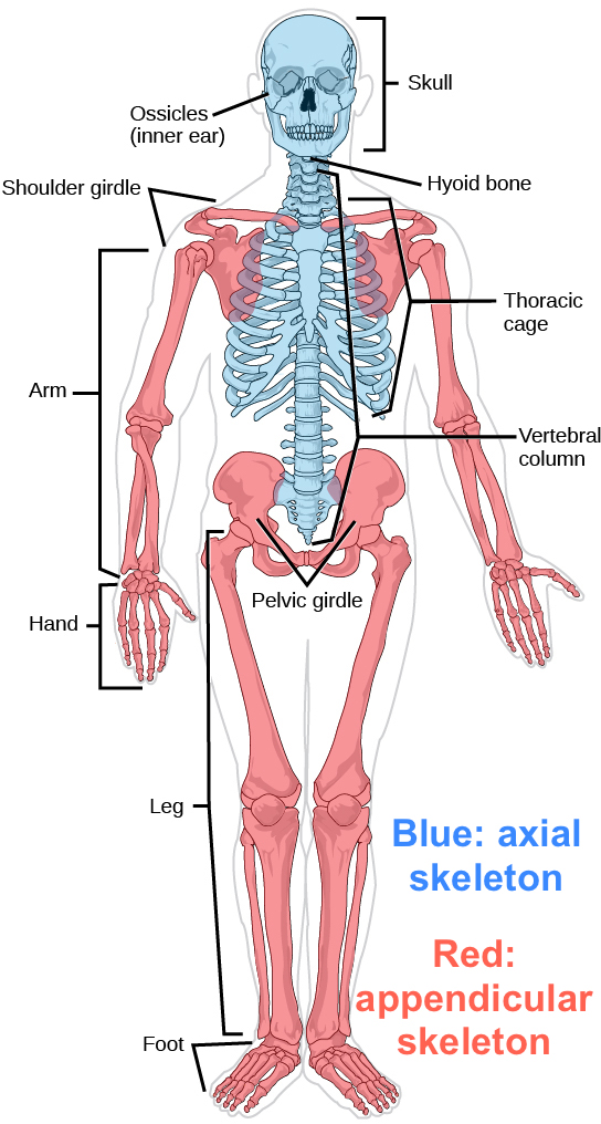
Labelled human skeleton with the axial skeleton shown in blue and the appendicular skeleton in red.

Figure from the supplied lecture notes: the axial skeleton (blue) is the central axis — skull, vertebral column and rib cage; the appendicular skeleton (red) is the limbs with their girdles.

Original figure labels are retained. Enlarge to inspect them; the explanation is available in the selected language.

Conceptual joint cutaway, not a named joint and not to scale. Blue cartilage coats both facing bone ends; the gap between them is the joint cavity. The fine outline is the capsule, the lower band a ligament, and the upper cords are tendons. Vessels, nerves and the opposing muscle are omitted.

Key points to remember

  • The musculoskeletal system has four jobs: support and shape, movement, protection of organs, and blood cell production in bone marrow.
  • Bone is living tissue: compact and cancellous tissue, four cell types, a periosteum that carries vessels and nerves, and marrow that makes blood cells.
  • Bones are classified as long, short, flat or irregular, and the skeleton is divided into the axial (about 80) and appendicular (about 126) parts.
  • Joints are immovable or movable; the more a joint moves, the more it depends on ligaments and capsule for stability, which is why sprains and dislocations happen there.
  • Skeletal muscle is voluntary, striated, attaches by tendon and can only pull; it therefore needs an opposing muscle, and it keeps pulling on bone fragments after a fracture.
Go deeper

Worked example

Reading a fracture description aloud, term by term. The handover sheet says: “Closed displaced oblique fracture of the mid-shaft of the right tibia. Neurovascular status intact.” Instead of memorising the sentence, use the structure and function you have just learned to unpack it.

  1. Name the bone and the region. The tibia is a long bone of the appendicular skeleton, in the lower limb, so it is a weight-bearing lever. Any loss of alignment therefore threatens the patient’s ability to stand, not just the appearance of the leg.
  2. Read the pattern. “Oblique” describes the direction of the break: it crosses the bone diagonally rather than straight across. The pattern tells you how stable the break is and whether the two ends are likely to slide past each other under load.
  3. Read open or closed. The word “closed” means the skin is intact, so the periosteum and the tissues around the bone were not exposed to the outside. This matters because the periosteum carries the blood vessels and nerves, so a closed fracture still bleeds and hurts inside, but it carries a much lower risk of infection than an open one.
  4. Read displacement and alignment. “Displaced” means the two ends are no longer in their normal position. This is exactly what the attached muscles do when they keep pulling after the break, which is why the limb is immobilised or placed in traction until the bone is stabilised.
  5. Read the safety statement. “Neurovascular status intact” tells you the nerves and blood vessels running beside the bone have not yet been compromised. It is the most important line for the nurse, because it is the finding that can change in the next hour and it is assessed by pulse, colour, warmth, sensation and movement distally.

Every unfamiliar fracture sentence can be unpacked in the same order: which bone and where, what pattern, open or closed, displaced or not, and what the neurovascular status is. Notice that only two of those questions come from the words on the page; the rest come from knowing that bone is a living, bleeding, load-bearing lever with muscles still pulling on it.

Common misconception

Many beginners think of bone as a dead, hard, unchanging scaffold, like the dry bones of a skeleton diagram. Bone is living tissue with a blood supply, its own nerves and cells that constantly build and remove it, which is why it bleeds and hurts when broken, why it remodels after a fracture, and why a hormonal or dietary change can make the whole skeleton weaker over months. Treating bone as inert makes fracture healing, osteoporosis and Paget’s disease impossible to understand.

Check your reasoning

A patient has a swollen, unstable knee after a twisting injury, and the X-ray shows no broken bone. Using the tissues you learned in this section, explain what has probably been injured and why the joint is unstable.

Reveal an answer

The likely injury is to the soft tissues that stabilise the joint rather than to bone. The knee is a freely movable joint, so its stability depends on the capsule and ligaments, not on interlocking bone. A twisting force that stretches or tears a ligament produces a sprain: the joint still looks intact on X-ray because the bone is unchanged, but it can no longer resist the movement the ligament normally limits, which is why it feels unstable and why it swells as the torn tissue bleeds into the joint. This is also why a normal X-ray never excludes a significant knee injury, and why the joint is examined for laxity rather than only for fracture.

Where this comes from

  • MUSKULOSKELETAL SYSTEM PATHOLOGIES lecture notes (supplied), Level 200 nursing · Lines 1–102: learning objectives, the “musculoskeletal system” and its terms table (joint, cartilage, ligament, voluntary/involuntary and striated muscle), functions of the skeleton, the axial (80 bones) and appendicular (126 bones) divisions, bone tissue types (compact, cancellous, subchondral) with the periosteum, the 206-bone count, bone-cell table (osteoblast, osteoclast, osteocyte, haematopoietic), classification of bones by shape, and the immovable/movable joint division.
  • Beyond the supplied notes: standard anatomy and physiology of bone growth and remodelling · the remodelling cycle and its coupling, the calcium and vitamin D control of bone turnover with the figure that about 99 per cent of body calcium is skeletal, intramembranous and endochondral ossification, the epiphyseal plate as the site of growth in length and its link to achondroplasia, and synovial joint anatomy (articular cartilage, synovial membrane and fluid, capsule, ligaments and bursae). The supplied notes list the bone cells and tissue types but do not explain the mechanisms, which are uncontroversial standard physiology.

Lesson progress is saved on this device for the current learner. Practice answers stay separate and are not stored.

About this course and its sources

The musculoskeletal and neurological lessons build on the supplied Level 200 nursing notes. Reproductive, respiratory, skin and nursing-response lessons use cited external references. Diagrams are original educational schematics. Sections that go beyond the supplied notes say so in their sources. Specialist clinical and French-language editorial review remain pending.