NU 237 · NursingSection 4.4 · Fluid balance and the urinary system
Section 12 of 25
Urinary retention, obstruction and hydronephrosis
Explain the difference between acute, chronic and acute-on-chronic urinary retention, list the obstructive, infective, mechanical, neurological and drug causes, trace how an obstruction backs urine up into the kidney to cause hydronephrosis, and describe the nursing care that relieves the obstruction and monitors the diuresis that follows.
New to this course? How each section works
Learn gives you one short idea at a time. Each idea starts with a single sentence, then explains it, gives an example, and asks you a small question you can answer straight away.
Explore shows a diagram of the same ideas. Click the numbered parts to read what each one is.
Check asks a few questions. Answer them all correctly to finish the section and unlock the next one.
“Go deeper” is optional. It adds mechanism and extra detail for when you are ready — nothing you need for the checks is hidden there.
Why this matters
A patient who cannot pass urine is in pain, and an obstruction that is left in place can quietly destroy a kidney. Retention and obstruction run through this whole unit: they are common, the nurse is usually the first person to recognise them, and the treatment — relieving the blockage and then watching the urine that follows — is a nursing task from start to finish. Learn the chain that runs from a narrowed outlet to a scarred kidney and you will recognise a distended bladder, ask for the right test, and understand why the next 24 hours of urine output can change the patient’s outcome.
What you need to know first
You need only the everyday picture that urine is made in the kidneys, travels down two tubes called ureters to the bladder and leaves through the urethra, and that an older man’s prostate sits around that outlet. Retention, residual volume, hydronephrosis and dyssynergia are all defined below, so no previous urology course is assumed.
Words to know (6)
Urinary retention
The inability to pass urine, which may come on suddenly with pain (acute) or develop slowly and painlessly over months (chronic). A man who has passed urine all his life suddenly cannot void at all and has a painful, swollen lower abdomen.
Post-void residual volume
The amount of urine still in the bladder immediately after the patient has passed urine, measured by a bedside bladder scan. A patient passes only a small trickle, yet the scan shows 600 mL still in the bladder, so the voiding was not complete.
Bladder outlet obstruction
A narrowing at or below the bladder neck that makes the bladder work harder to empty; an enlarged prostate is the commonest cause in older men. Benign prostatic hyperplasia narrows the urethra, so the bladder empties incompletely and residual urine accumulates.
Hydronephrosis
Swelling of the kidney and its collecting area caused by urine backing up behind an obstruction, which raises the pressure inside the kidney. An ultrasound shows a swollen left kidney with a dilated renal pelvis in a man whose bladder no longer empties.
Detrusor–sphincter dyssynergia
A loss of coordination in which the bladder muscle contracts while the urethral sphincter stays closed, so the bladder squeezes against a shut outlet. In spinal cord injury the detrusor contracts but the sphincter does not relax, producing high pressures and incomplete emptying.
Post-obstructive diuresis
A large, sometimes excessive production of urine in the hours after a long-standing obstruction is relieved, because the kidney has lost its ability to concentrate urine. After 1.4 litres are drained from a chronically distended bladder, the patient passes more than 200 mL of urine every hour.
One idea at a timeIdea 1 / 6
In one sentence Urinary retention means the patient cannot pass urine; acute retention is sudden and painful, while chronic retention builds up slowly and may hardly hurt.
Retention is the inability to pass urine, and it may be acute, chronic or both
Retention is an inability to pass urine, and the two forms behave differently. Acute urinary retention is a new-onset inability to pass urine, which then causes pain and discomfort together with significant residual volumes. One detail matters in practice: some patients still pass small quantities of urine yet keep significant residual volumes and ongoing discomfort, so they are still in acute retention. Chronic retention develops gradually, the bladder never empties properly, and the patient may tolerate it with little pain. Retention is most prevalent in older men, usually because an enlarged prostate obstructs the bladder outlet, but the causes are many. A third pattern is acute-on-chronic retention: a patient with chronic retention suddenly deteriorates, either because the underlying disease worsens or because a new problem is added. These patients often have minimal discomfort despite very large residual volumes, and after treatment they are more at risk of post-obstructive diuresis than other patients in acute retention.
For example
An 82-year-old man cannot pass urine at all since waking, and his lower abdomen is tense and painful. A younger man in chronic retention may carry a litre of residual urine for weeks with only a dull sensation.
Pause and check yourself
Why does a patient who is still passing a little urine sometimes have acute retention?
Show the answer
Because the amount passed is not the deciding feature: if the bladder is not emptying, residual volume and discomfort remain, and that is acute retention however small the stream.
Read detailed explanations
The pressure cascade: from a blocked outlet to a scarred kidney
Obstruction damages the kidney through a sequence of pressures, and each step can be measured. Urine is normally propelled by gentle peristaltic waves that generate only a few centimetres of water of pressure inside the renal pelvis, so the tubules downstream are never exposed to a significant hydrostatic load. When an obstruction appears, whether it is a prostatic enlargement, a calculus or a fibrous stricture, the fluid behind it becomes static. Because the tract is a closed space, the pressure generated by the collecting system is transmitted backwards, rising first in the ureter and then in the renal pelvis. The kidney responds with distension, which is hydronephrosis, but the visible swelling matters less than the pressure it represents, because that pressure is transmitted to the nephrons themselves. Within hours of an acute complete obstruction, the glomerular filtration rate falls substantially; the cause is a combination of reduced renal blood flow and a rising hydrostatic pressure in Bowman’s space that opposes filtration. Tubular function is impaired at the same time: the ability to transport sodium, potassium and protons, and to dilute or concentrate the urine, is severely reduced, so the kidney loses the fine control that keeps the internal environment steady. If the obstruction is relieved quickly, much of this recovers, but the depression of glomerular filtration can persist for weeks, and a kidney that has been subjected to repeated or prolonged episodes develops interstitial fibrosis as the muscular wall of the ureter is replaced by collagenous scar tissue and the renal parenchyma thins through pressure atrophy. This is why repeated high-pressure episodes cause permanent scarring and chronic kidney disease. Bilateral obstruction is far more dangerous than unilateral, because the unobstructed contralateral kidney can compensate for one damaged organ but not for two, and it is bilateral obstruction that produces azotaemia and, if untreated, uraemic complications.
Post-obstructive diuresis: the numbers to watch after the tap is opened
Relieving a long-standing obstruction does not always end the problem. The distended kidney has been building and maintaining a salty medullary gradient so that it can reabsorb water from the collecting duct; when the pressure is suddenly removed, that gradient is washed out and the tubule can no longer conserve water. At the same time, the urea and sodium that accumulated during the obstruction are delivered to the tubule in large amounts and act as an osmotic diuretic, dragging water out with them. The result is post-obstructive diuresis: a large volume of dilute urine that can continue for hours to days. The clinical risk is that this fluid is lost from a patient who is already unwell, so plasma volume falls, electrolyte reserves are depleted, and the acute kidney injury that the catheter was meant to prevent can worsen. There is a clear threshold for action: patients producing more than 200 mL of urine per hour should have about half of that output replaced with intravenous fluid, and their urine output is monitored over the 24 hours after catheterisation. Not every patient needs this. The risk is concentrated in those with a very large residual volume, and volumes above about 1000 mL are seen only in acute-on-chronic retention, which is why the drained volume is recorded and not simply discarded. Nursing observation therefore has three parts: an hourly measurement rather than a 24-hour total, because the threshold is expressed per hour; a record of the colour and concentration, since dilute urine confirms the mechanism; and regular monitoring of renal function and electrolytes, particularly potassium and magnesium, because replacement fluid restores volume but not necessarily the solutes lost with it. Patients whose function is still impaired also keep their catheter, since a further retention episode would add damage on top of damage.
Obstruction across the lifespan: why the cause changes with age
The site of an obstruction is often predictable from the patient’s age, and that pattern is a useful first question. In children, obstruction is due mainly to birth defects, and the classic example is posterior urethral valves, extra folds of tissue in the back part of the urethra that narrow the outflow before the child is ever toilet trained; other congenital constrictions narrow the ureter or the junction between the ureter and the renal pelvis. In young adults the commonest cause is a stone lodged in a kidney, a ureter or elsewhere, and because stones are mobile and irregular they typically produce the intermittent agony of renal colic rather than the silent distension of a slow obstruction. In older adults the balance shifts to benign prostatic hyperplasia and prostate cancer, together with tumours and stones, which is why obstruction is more common in men than in women overall. Beyond that age pattern the list is long: polyps in the ureter, a blood clot, a tumour pressing on the ureter from outside, disorders of the muscle or nerve of the ureter or bladder, fibrous tissue forming around the ureter after surgery, radiation therapy or certain drugs such as methysergide, a ureterocele, which is a bulging of the lower ureter into the bladder, tumours, abscesses and cysts of the bladder, cervix, uterus, prostate or other pelvic organs, and a large mass of faeces stuck in the rectum. In women two mechanisms are particular to them: as the uterus expands in pregnancy it can compress the ureters, and a prolapsed uterus or a cystocele, in which the bladder droops because the wall between bladder and vagina has weakened, can distort the outflow. The practical message is that obstruction in a woman or a child is not rare and is not always prostatic, so the assessment follows the anatomy rather than the stereotype.
Explore the diagram
Look at the whole drawing first. Then select one numbered part and connect it to the idea you just read.
From a blocked outlet to a damaged kidney
Follow the pressure, not the anatomy. A narrowing at the bladder outlet makes the bladder empty incompletely, so residual urine accumulates and the pressure inside rises. If that pressure is high enough, it defeats the anti-reflux mechanism where the ureters enter the bladder, and urine is pushed backwards: the ureter dilates, the renal pelvis and kidney distend (hydronephrosis), and filtration and tubular work fall. Each step is something the nurse can detect and act on.
New to diagrams? Read this first
Look at the whole drawing once without clicking anything. Ask yourself only: what kind of picture is this (a comparison, a route, a sequence, a table)?
Press Play, or use the arrows, to move through the numbered parts in order. The drawing stays still; only the highlight moves.
Click a number, or its name in the list below, to read what that part is and why it matters. Use “Hide labels” when you want to test your memory.
More controls
01 / 5Bladder outlet obstruction
If the drawing is wider than this panel, scroll it sideways. Labels stay readable; the numbered explanations below show the same ideas.
Bladder outlet obstruction
The obstruction may sit inside the urethra, as in a stricture, or outside it, as when an enlarged prostate or a pelvic mass compresses the outflow. Whatever the cause, the bladder must generate more pressure to push urine past it, and that extra pressure starts the whole chain of damage. In older men benign prostatic hyperplasia is the commonest cause, while a stone, a clot, a tumour or a narrowed ureteric junction can obstruct at a higher level.
Supporting illustrations
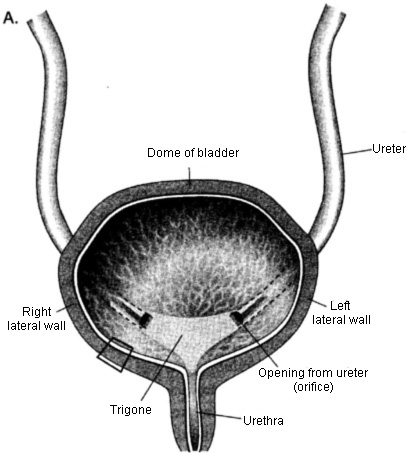
Figure from the supplied lecture notes: the landmarks used to say where a bladder problem sits, and the wall that holds the urine in.
Original figure labels are retained. Enlarge to inspect them; the explanation is available in the selected language.
Schematic chain of pressure, not an anatomical drawing: no organ is drawn, only a single symbol beside step 2 and the sequence of pressure steps as text boxes, placed in order rather than to scale. It shows the sequence of events and what the nurse can measure at each step, not the position of the organs.
Key points to remember
✓Retention is the inability to pass urine; acute retention is sudden and painful with significant residual volumes, chronic retention is slow and often painless, and acute-on-chronic retention may cause little discomfort despite a very large volume.
✓Causes are obstructive (enlarged prostate, stricture, prostate cancer), infective, mechanical (constipation), painful, pharmacological (antimuscarinics, spinal or epidural anaesthesia) and neurological (neuropathy, pelvic surgery, multiple sclerosis, Parkinson’s disease, detrusor–sphincter dyssynergia).
✓Obstruction anywhere from the kidney to the urethra raises the pressure behind it; urine backs up into the renal pelvis and distends the kidney, which is hydronephrosis, and stagnant urine favours stones and infection.
✓High-pressure retention overcomes the anti-reflux mechanism, so urine backs up into both kidneys, renal function falls and repeated episodes scar the kidney; low-pressure retention leaves the upper tract intact.
✓Treatment is immediate catheterisation with measurement of the drained volume, treatment of the cause, a trial without catheter when renal function is normal, and monitoring for post-obstructive diuresis, infection and kidney injury.
Go deeper
Worked example
An 84-year-old man with acute-on-chronic retention. An 84-year-old man with a long history of a poor stream is brought in after two days of being unable to pass urine. He says the discomfort is “not too bad”. His lower abdomen is distended but only mildly tender, and a bladder scan reads over 1,200 mL. His blood results show a creatinine of 210 micromol/L, well above his previous 95, and an ultrasound shows bilateral hydronephrosis.
Recognise the pattern. The long history of a poor stream is chronic retention, and the sudden inability to pass urine is the acute event on top of it. The mismatch between a very large residual volume and only mild discomfort is typical of acute-on-chronic retention.
Relieve the obstruction and measure what comes out. Immediate urethral catheterisation drains the bladder; the drained volume is recorded because a drained volume above about 1000 mL is seen only in acute-on-chronic retention and signals a higher risk of post-obstructive diuresis.
Explain the abnormal blood results. The raised creatinine and bilateral hydronephrosis say that the bladder pressure had overcome the anti-reflux mechanism and backed urine up into both kidneys: this is high-pressure retention, not a kidney disease of its own.
Monitor for post-obstructive diuresis. Hourly urine output is charted for the next 24 hours. If the patient produces more than 200 mL per hour, the medical team prescribes intravenous fluid to replace roughly half of that output, because the kidney, having lost its ability to concentrate urine, can otherwise dehydrate the patient and worsen the kidney injury.
Plan the next steps and prevent recurrence. Because renal function is impaired, the catheter stays in place until definitive treatment of the prostate is arranged, and the urine specimen is sent to check for infection. A trial without catheter is not attempted while the kidney function is still recovering.
This patient’s kidneys were damaged by back-pressure, not by intrinsic kidney disease. Two nursing numbers change the outcome: the volume drained at catheterisation, which flags the risk, and the hourly urine output afterwards, which detects post-obstructive diuresis early enough to replace the fluid.
Common misconception
A very common beginner belief is that a patient who is passing some urine cannot be in retention, and that a falling urine output when the bladder is full means the kidneys have stopped making urine. Neither is true. Patients may pass small quantities while still retaining a significant residual volume and discomfort, so a trickle of urine never excludes retention, and the bladder scan, not the patient’s description, settles the question. In obstruction the kidney is usually still filtering: the urine is produced and then trapped, which is why draining the bladder can release a litre or more at once.
Check your reasoning
A patient with chronic retention has 1.3 litres drained at catheterisation. Over the next six hours he passes 1.5 litres. Using this section, explain what is happening and what you must do.
Reveal an answer
The large drained volume identifies acute-on-chronic retention and warns of post-obstructive diuresis. The kidney has lost the medullary concentration gradient it needs to conserve water, and the retained urea and sodium act as an osmotic diuretic, so it produces a large volume of dilute urine. Because losing this fluid can worsen kidney injury and deplete the circulation and electrolytes, the output is measured hourly and roughly half of it is replaced with intravenous fluid as prescribed, with regular checks of renal function and electrolytes.
Where this comes from
Urinary System Pathologies lecture notes (supplied), Level 200 nursing · “Introduction” and “Aetiology”, lines 88–140: acute, chronic and acute-on-chronic retention, the definition with significant residual volumes, the note that some patients still pass small quantities of urine, the aetiology list (benign prostatic hyperplasia, urethral strictures, prostate cancer, urinary tract infection, constipation, severe pain, antimuscarinics and spinal or epidural anaesthesia, neuropathy, pelvic surgery, multiple sclerosis and Parkinson’s disease, bladder–sphincter dyssynergia), clinical features and investigations, high-pressure retention with hydroureter and hydronephrosis, management including tamsulosin and the trial without catheter at 24–48 hours, the arbitrarily >1000 mL drainage volume seen only in acute-on-chronic retention, post-obstructive diuresis with the >200 mL/hour threshold and the 50 per cent replacement rule, and complications. “Urinary tract obstruction” and “Hydronephrosis”, lines 607–732: causes by age group, symptoms including renal colic, diagnosis, relief of obstruction, hydronephrosis definitions, the pathophysiology with the fall in glomerular filtration rate and impaired tubular transport, clinical manifestations and treatment.
Beyond the supplied notes: standard urological and renal knowledge, with corrections · the oblique entry of the ureter and the vesicoureteral anti-reflux mechanism, the opposition of filtration by hydrostatic pressure in Bowman’s space, the loss of the medullary concentration gradient with the osmotic action of retained urea in post-obstructive diuresis, the monitoring of potassium and magnesium after large-volume replacement, and posterior urethral valves as the classic paediatric cause of obstruction. Corrections to the supplied notes: “Uterocele” is corrected to ureterocele, “uremi” to uraemia and “Pruritis” to pruritus; the unitless “large residual volume” is tied to the source’s own figure of around >1000 mL, which the notes state occurs only in acute-on-chronic retention. The supplied notes’ nursing-care plan for hydronephrosis lists “risk for infection related to hemodialysis access”, which does not apply to a patient with hydronephrosis; this is corrected here to the risk of infection related to urinary stasis and obstruction.
Lesson progress is saved on this device for the current learner. Practice answers stay separate and are not stored.
About this course and its sources
The urinary and endocrine lessons build on the supplied Level 200 nursing notes (course instructor Bakume Jessey). Disease mechanisms, blood, lymphatic, cardiovascular and digestive lessons use cited external references. Diagrams are original educational schematics. Sections that go beyond the supplied notes say so in their sources. Specialist clinical and French-language editorial review remain pending.