← Common Core coursesNU 237 · Medical Pathology I
NU 237 · NursingSection 4.1 · Fluid balance and the urinary system
Section 9 of 25

How the kidney controls fluid, electrolytes and acid–base balance

Explain what the urinary system is for, how the nephron filters and then adjusts the blood, and how the kidney keeps water, electrolytes and pH inside the narrow range that cells tolerate.

New to this course? How each section works
  1. Learn gives you one short idea at a time. Each idea starts with a single sentence, then explains it, gives an example, and asks you a small question you can answer straight away.
  2. Explore shows a diagram of the same ideas. Click the numbered parts to read what each one is.
  3. Check asks a few questions. Answer them all correctly to finish the section and unlock the next one.
  4. “Go deeper” is optional. It adds mechanism and extra detail for when you are ready — nothing you need for the checks is hidden there.
Why this matters

Almost every medical condition in this unit is, at bottom, a disturbance of what the kidney is supposed to hold steady. Retention, infection, glomerular disease, stones and kidney failure all end in the same language: too little or too much water, a potassium that can stop the heart, a pH that paralyses enzymes. If you understand the three jobs of the nephron now, the signs you will meet later in the unit stop being a list to memorise and become a prediction you can make at the bedside. This section is the physiology that the rest of the urinary unit depends on.

What you need to know first

You need only the everyday idea that the body is mostly water, that cells need the fluid around them to stay roughly the same, and that the kidneys produce urine. The words nephron, filtration and reabsorption are defined below, so no previous physiology course is assumed.

Words to know (5)
Urinary system
The two kidneys, two ureters, bladder and urethra, which together filter blood and carry the waste away as urine. A blocked ureter stops urine reaching the bladder even though the kidney still makes it.
Nephron
The microscopic working unit of the kidney: a ball of capillaries called the glomerulus joined to a tube, the renal tubule. Each kidney holds about a million nephrons, and each one makes its own drop of urine.
Glomerular filtration
The first step of urine formation, in which blood pressure pushes water and small dissolved substances out of the glomerulus into the tubule. If blood pressure falls far enough, filtration falls too and less urine is made.
Reabsorption
The process by which the tubule takes useful filtered substances back into the blood instead of losing them in the urine. Glucose is normally reabsorbed completely, so healthy urine contains no sugar.
Acid–base balance
The control of the acidity of body fluids, measured as pH, so that enzymes and cell membranes keep working. When the kidneys fail, acid accumulates and the patient breathes deeply to compensate.
One idea at a timeIdea 1 / 5

In one sentence The urinary system filters blood, removes waste as urine, and keeps water, salts and pH inside the narrow range that cells need.

The urinary system removes waste and holds the internal environment steady

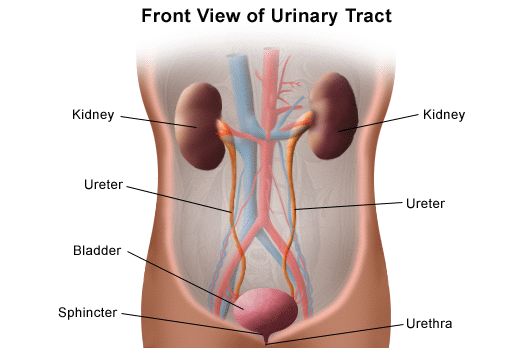
Two kidneys sit below the ribs toward the middle of the back. Each contains about a million nephrons, and each nephron is a filter joined to a tube. Blood arrives under pressure, water and small dissolved substances are pushed out, and the tube then decides what to keep and what to let go. What leaves the kidney travels down a ureter to the bladder, a hollow muscular bag that stores urine until the brain signals the sphincters to relax. The kidney is therefore both a waste-disposal organ and the controller of the fluid around every cell: it removes urea, drugs and excess ions, and at the same time holds volume, electrolyte concentration and pH steady. Normal urine is pale straw-coloured; darker urine usually means the body is conserving water.

For example

A patient who drinks nothing for a day still passes urine, but it is small in volume and dark. The kidney has kept the waste flowing while conserving water.

Pause and check yourself

Name the two jobs of the urinary system in one sentence.

Show the answer

It removes waste products from the blood as urine, and it keeps the volume, electrolyte concentration and pH of body fluids steady.

Read detailed explanations
How the tubule concentrates urine: the loop of Henle and the hormones that follow it

Reabsorption alone would leave urine roughly as concentrated as blood. To make urine several times more concentrated, or several times more dilute, the nephron uses an arrangement called the countercurrent multiplier. The loop of Henle dips deep into the medulla of the kidney, the inner zone around the renal pelvis that the rest of the nephron does not enter. Its descending limb is permeable to water but not to salt, so as fluid descends into the salty medulla, water leaves by osmosis and the fluid inside becomes more concentrated. Its ascending limb is the opposite — impermeable to water but actively pumping sodium and chloride out — so the fluid becomes dilute as it climbs, while the pumped salt builds and maintains the medullary gradient. Because the two limbs run side by side in opposite directions, each pass amplifies the gradient rather than washing it out. The vasa recta, hairpin capillaries that follow the loop, carry the reabsorbed water away without destroying the gradient. The result is a medulla that becomes progressively saltier toward the papilla, creating the osmotic force that can pull water out of the collecting duct.

Whether that force is used depends on vasopressin, also called antidiuretic hormone, released from the posterior pituitary. Vasopressin inserts water channels called aquaporin-2 into the collecting duct wall, so water follows the gradient back into the blood and a small volume of concentrated urine is produced. When vasopressin is suppressed, the channels are withdrawn, the collecting duct stays watertight, and a large volume of dilute urine is passed. A second hormone, aldosterone from the adrenal cortex, acts further upstream in the distal tubule, where it increases sodium reabsorption and potassium secretion; because water follows the reabsorbed sodium, aldosterone effectively expands the circulating volume. Aldosterone is the final step of the renin–angiotensin–aldosterone system: low renal perfusion pressure, low sodium delivery or sympathetic stimulation makes the kidney release renin, which converts angiotensinogen to angiotensin I, which angiotensin-converting enzyme converts to angiotensin II, a potent vasoconstrictor that also triggers aldosterone release.

Clinically these pieces explain concrete findings. A patient with a high vasopressin level, as in the syndrome of inappropriate antidiuretic hormone secretion, retains water and becomes hyponatraemic with concentrated urine. A patient with no vasopressin, as in cranial diabetes insipidus, passes many litres of dilute urine and becomes hypernatraemic if fluids are withheld. A patient on a loop diuretic has that medullary gradient itself dismantled, which is why loop diuretics are powerful and also why they waste potassium and can disturb calcium and magnesium handling.

Reading the numbers: what each fluid, electrolyte and acid–base result means

Because the kidney sets the composition of body fluids, the laboratory report is really a report on the tubule. A useful approach is to read four things together rather than one at a time. First, the effective circulating volume: daily weight, intake and output, pulse, blood pressure, mucous membranes and skin turgor. Second, the sodium concentration, normally about 135 to 145 mmol/L. Sodium is not a measure of how much salt the body holds but of how much water dilutes it. A low sodium usually means relatively too much water, which can happen with heart failure, liver disease, or inappropriate vasopressin, and not only with salt loss. A high sodium almost always means a water deficit relative to salt.

Third, potassium, normally about 3.5 to 5.0 mmol/L. Potassium matters out of proportion to its number because it governs the resting electrical potential of every cell, especially cardiac muscle: a low value causes weakness, cramps and arrhythmias, while a high value causes muscle weakness, paraesthesia and, above roughly 6.5 mmol/L, classic electrocardiogram changes — tall peaked T waves, then a widened QRS, then a sine-wave pattern that precedes cardiac arrest. Fourth, the acid–base picture, usually read from a blood gas, with bicarbonate normally about 22 to 26 mmol/L and arterial pH about 7.35 to 7.40. The bicarbonate tells you the metabolic side and the carbon dioxide the respiratory side; the two normally move in opposite directions as compensation. When the bicarbonate is low with a low pH, the patient has metabolic acidosis, and the cause is found by considering the anion gap, the difference between measured cations and anions. A normal-gap acidosis, as in diarrhoea, is bicarbonate lost from the gut. A raised-gap acidosis, as in lactic acidosis, ketoacidosis or advanced kidney failure, is acid added to the body. When the bicarbonate is high with a high pH, the patient has metabolic alkalosis, as in prolonged vomiting with loss of stomach acid.

Two quantitative rules are worth carrying to the bedside. The first is that urine output below about 0.5 mL per kilogram per hour in an adult is oliguria and needs explanation. The second is that a serum creatinine rise of as little as 26 micromol/L in 48 hours already meets the definition of acute kidney injury, because creatinine is a late and insensitive marker and the tubule has usually lost much of its reserve before the number moves. Finally, the concentration of the urine itself is informative: a dilute urine in a patient who looks dry suggests the tubule has lost its concentrating ability, whereas a concentrated urine in the same patient suggests the kidney is working hard to conserve.

When regulation fails: what the nurse monitors and why

Nursing monitoring of a renal patient is not a ritual of recording numbers; each number tests one of the kidney’s jobs, and the value of the measurement lies in the decision it triggers. Daily weight is the most sensitive bedside index of fluid balance because a litre of retained water weighs a kilogram, while oedema and even lung crepitations appear later. Strict intake and output, measured on every shift, detect a falling urine output before the blood chemistry changes, and the character of the urine — colour, cloudiness, sediment — points to concentration, infection or stones. Blood pressure is monitored because the kidney both controls it and is damaged by it, and because a rising pressure may be the first sign of fluid overload.

Serum potassium deserves specific attention because it is the electrolyte that kills without warning: the nurse must know which patients are at risk, including those in kidney failure, those on angiotensin-converting enzyme inhibitors, potassium-sparing diuretics or potassium supplements, and those with diabetes or metabolic acidosis. Because the electrocardiogram changes of hyperkalaemia progress in a predictable order, a patient with a suspiciously high potassium is placed on continuous cardiac monitoring while treatment is prepared. Fluid overload is recognised by weighing the patient, listening to the lung bases, checking jugular venous pressure and looking for pitting oedema, and it is treated by restricting fluid and sodium and by giving the prescribed diuretic at the time that best matches the patient’s pattern of urine production. Uraemia, the syndrome of retained waste products, produces a characteristic cluster the nurse charts: nausea and vomiting, anorexia, a metallic taste, itching, drowsiness, confusion and eventually a pericardial rub or seizure. Drug therapy is monitored differently in kidney disease because the kidney is the main elimination route for many drugs: doses are reduced, dosing intervals extended, and levels checked, and the nurse who gives an aminoglycoside or a renally cleared opioid without checking the renal function is creating the toxicity she will later have to report.

Patient education follows the same logic. The patient is taught which foods carry potassium and phosphate, why protein and sodium may be restricted, why fluid may be limited to a prescribed volume, how to recognise the warning symptoms of hyperkalaemia or fluid overload, and why every non-prescribed medicine, including herbal remedies and painkillers bought without prescription, must be checked with the health worker first. Finally, the nurse watches the access route: a fistula or a peritoneal dialysis catheter is the patient’s lifeline, so its thrill, bruit, cleanliness and the absence of pressure on that arm are assessed every shift, and any suspicion of infection or loss of function is escalated immediately rather than documented and left.
Explore the diagram

Look at the whole drawing first. Then select one numbered part and connect it to the idea you just read.

The nephron: filter first, reclaim afterwards

Follow one drop of fluid through the working unit of the kidney. Blood arrives under pressure; the glomerulus filters water and small solutes out; the tubule reabsorbs almost all of what the body still needs; the collecting duct makes the final hormone-controlled adjustment; what is left becomes urine. The peritubular capillary carries the reclaimed material back to the blood, while regulated amounts of water and solutes are excreted.

New to diagrams? Read this first
  1. Look at the whole drawing once without clicking anything. Ask yourself only: what kind of picture is this (a comparison, a route, a sequence, a table)?
  2. Press Play, or use the arrows, to move through the numbered parts in order. The drawing stays still; only the highlight moves.
  3. Click a number, or its name in the list below, to read what that part is and why it matters. Use “Hide labels” when you want to test your memory.
More controls
100%

01 / 4Glomerulus: filtration under pressure

The nephron: filter first, reclaim afterwardsFollow one drop of fluid through the working unit of the kidney. Blood arrives under pressure; the glomerulus filters water and small solutes out; the tubule reabsorbs almost all of what the body still needs; the collecting duct makes the final hormone-controlled adjustment; what is left becomes urine. The peritubular capillary carries the reclaimed material back to the blood, while regulated amounts of water and solutes are excreted.CORTEX · filtration and early recoveryMEDULLA · concentration gradientBlood inBlood leaves the glomerulusTubule → bloodWater and useful solutesSecretion: blood → tubuleLoop of HenleUrine → renal pelvisRed route: blood · blue tube: filtrate and urineArrows show flow or transfer; thin leaders identify structures.

If the drawing is wider than this panel, scroll it sideways. Labels stay readable; the numbered explanations below show the same ideas.

Glomerulus: filtration under pressure

The glomerulus is a knot of capillaries where blood pressure pushes water and small dissolved substances through a fine filter into the tubule. Because the process is driven by pressure, anything that lowers renal perfusion — shock, severe dehydration, heart failure — lowers the volume filtered each minute. Blood cells and large proteins are held back, heavy protein loss may indicate filter injury, but blood in urine can also come from elsewhere in the urinary tract.

Supporting illustrations

1 / 3
Labelled front view of the urinary tract: two kidneys, two ureters, the bladder, a sphincter and the urethra.

Figure from the supplied lecture notes: the organs this section describes, in the order urine travels — kidney, ureter, bladder, sphincter, urethra.

Original figure labels are retained. Enlarge to inspect them; the explanation is available in the selected language.

Schematic of one nephron, simplified: the true tubule is far longer and folds many times. The red return route stands for a capillary network; medullary vessels and neighbouring nephrons are omitted. Collecting ducts receive fluid from several nephrons. Positions and proportions are schematic.

Key points to remember

  • The urinary system has two jobs at once: it removes waste as urine and it keeps fluid volume, electrolyte concentration and pH steady.
  • The nephron filters under pressure first, then reabsorbs what the body still needs and secretes a few substances on purpose; urine is what remains.
  • Water follows sodium, so the kidney never manages one without the other, and a fluid problem is described by both the volume lost and the sodium concentration.
  • The lungs correct pH within minutes but only for carbon dioxide; the kidneys correct it completely over hours by excreting acid and regenerating bicarbonate.
  • The kidney also makes erythropoietin, releases renin, activates vitamin D and clears many drugs — so kidney failure causes anaemia, hypertension, bone disease and drug accumulation.
Go deeper

Worked example

A patient vomiting for two days: what the kidney is doing. A 45-year-old woman has vomited repeatedly for two days and cannot keep fluids down. She is thirsty, her pulse is 104 beats per minute, her blood pressure has fallen from 120/80 to 100/65 mmHg, and her urine output is small and dark. Her blood results show a raised creatinine and a low bicarbonate.

  1. Identify the losses. Vomiting removes water, sodium, potassium and stomach acid. The patient has lost fluid from the body and acid from the stomach at the same time.
  2. Follow the volume signal. The falling blood volume is sensed by the kidney, which releases renin and starts the hormone chain that makes the tubule retain sodium and water. This is why the urine becomes small and dark: the kidney is conserving, not failing to produce.
  3. Follow the pressure signal. Because filtration depends on pressure, the drop from 120/80 to 100/65 mmHg lowers the volume filtered each minute. Less fluid is filtered, so less urine is made and creatinine rises in the blood.
  4. Follow the acid signal. Two forces push pH in opposite directions here. Loss of stomach acid with the vomit removes acid from the body, but the low blood volume also underperfuses the tissues, and poorly perfused tissue makes lactic acid. The low bicarbonate tells you the acid load is winning and the buffer reserve is being used up. The lungs answer by breathing more deeply and rapidly to blow off carbon dioxide — the deep breathing you will see in a patient with metabolic acidosis.

The dark, small volume of urine is the key bedside clue: it tells you the kidney is conserving fluid because the body is short. The raised creatinine does not yet prove permanent kidney damage, because part of it is simply reduced filtration from low pressure. Restoring the circulating volume with the prescribed fluid should restore urine output and bring the creatinine down — which is exactly what the nurse watches for by measuring intake, output and daily weight.

Common misconception

A common beginner belief is that urine is simply “leftover water” that the body did not need, so drinking more must always be good. In fact the kidney manufactures urine of a chosen concentration: it can put out a litre of dilute urine to shed excess water, or a few hundred millilitres of concentrated urine to defend the blood volume. That is why a patient can be overhydrated with a normal urine volume, and why a falling urine output is a warning about the circulation rather than a simple record of how much was drunk.

Check your reasoning

A patient’s urine output falls to 300 mL in 24 hours. Before you look at any blood result, what three questions does this section teach you to ask?

Reveal an answer

Ask first about the circulation: is the blood pressure or blood volume low enough to reduce filtration, and is the kidney conserving water because the body is short? Ask second about the outflow: is urine being made but unable to leave, as in obstruction? Ask third about the kidney tissue itself: is the tubule damaged, which would show as a loss of the substances it should be reabsorbing? Only then do the blood results tell you which of the three is most likely.

Where this comes from

  • Urinary System Pathologies lecture notes (supplied), Level 200 nursing · “Review of the Urinary System”, lines 1–38: the four organs, the nephron as a glomerulus joined to a renal tubule, urine colour and the kidney’s roles in blood pressure, erythropoietin and acid–base balance; “Urinary retention”, lines 88–140, for the effect of low perfusion and post-obstructive diuresis on urine output.
  • Beyond the supplied notes: standard renal physiology and reference ranges · countercurrent multiplication and the medullary gradient, aquaporin-2 and vasopressin action, the renin–angiotensin–aldosterone sequence, the definition of oliguria (0.5 mL/kg/h) and of acute kidney injury (a creatinine rise of 26 micromol/L in 48 hours), and reference ranges for sodium, potassium, bicarbonate, arterial pH and urine osmolality. These are uncontroversial standard values the supplied notes do not state.

Lesson progress is saved on this device for the current learner. Practice answers stay separate and are not stored.

About this course and its sources

The urinary and endocrine lessons build on the supplied Level 200 nursing notes (course instructor Bakume Jessey). Disease mechanisms, blood, lymphatic, cardiovascular and digestive lessons use cited external references. Diagrams are original educational schematics. Sections that go beyond the supplied notes say so in their sources. Specialist clinical and French-language editorial review remain pending.