BMS 114 · Common Core (Tronc commun)Section 1.2 · The microbial world
Section 2 of 33
The major groups of microorganisms
Distinguish the five groups selected for this section by cell organisation and replication, and explain the limits of identifying microbes from appearance or failed culture.
New to this course? How each section works
Learn gives you one short idea at a time. Each idea starts with a single sentence, then explains it, gives an example, and asks you a small question you can answer straight away.
Explore shows a diagram of the same ideas. Click the numbered parts to read what each one is.
Check asks a few questions. Answer them all correctly to finish the section and unlock the next one.
“Go deeper” is optional. It adds mechanism and extra detail for when you are ready — nothing you need for the checks is hidden there.
Why this matters
A microbe’s organisation helps explain how it grows and which tests or medicines may be useful. Bacteria and archaea have no membrane-bound nucleus, fungi and protozoa are eukaryotes, and viruses are acellular. These distinctions guide further investigation; they do not by themselves establish a diagnosis or prescription.
What you need to know first
You need no laboratory experience. The section uses one ordinary idea — a cell with or without a nucleus — and defines four technical words: prokaryote, eukaryote, binary fission and acellular. Read the key terms first if biology is new to you; the ideas that follow then add one group at a time, and the plate compares all five side by side.
Words to know (6)
Prokaryote
A cell whose genetic material is not enclosed in a true nucleus and which has no membrane-bound organelles. Bacteria and archaea are prokaryotes.
Eukaryote
A cell that has a true nucleus containing the genetic material, together with membrane-bound organelles. Fungi and protozoa are eukaryotes, and so are human cells.
Binary fission
Asexual division in which a cell copies its genetic material and divides into two daughter cells; mutations can introduce differences. Bacteria multiply by binary fission.
Acellular
Not organised as a cell: lacking cytoplasm and ribosomes. A virus may have a lipid envelope, but that envelope is not a complete cell. Viruses are the acellular agents in this five-group comparison.
Obligate intracellular replication
Dependence on a living host cell to make new infectious particles; viruses use host machinery rather than dividing as cells. Viruses infect animals, plants and microorganisms, including bacteria and archaea.
Mycelium and hyphae
The filamentous body of a mould: mycelium is the whole mass, and each individual branching thread is a hypha. The grey fuzz on old bread is mycelium made of hyphae.
One idea at a timeIdea 1 / 6
In one sentence This section compares bacteria, archaea, fungi, protozoa and viruses; these five groups are not an exhaustive classification.
Five selected groups: a useful comparison, not the whole field
Start with organisation: is the agent cellular or acellular? If cellular, does it have a membrane-bound nucleus? These questions distinguish viruses from cells, and prokaryotes from eukaryotes.
The supplied notes list bacteria, archaea, fungi, protozoa and viruses. We compare those five here, without claiming that all microbiology fits into five boxes. Microscopic algae and parasitic worms also belong within the wider field. These teaching groups are not equivalent taxonomic ranks; evolutionary relationships use molecular evidence as well as structure and activity.
For example
A pond contains bacteria, protozoa and microscopic algae. The algae do not stop being microbiological subjects because this particular comparison focuses on five other groups.
Pause and check yourself
Does comparing five selected groups mean that microscopic algae are excluded from microbiology?
Show the answer
No. The five groups define this section’s scope, not the boundaries of microbiology. Microscopic algae and parasitic worms are also studied, and viruses are compared separately because they are acellular.
Read detailed explanations
Selective toxicity: useful targets, not guaranteed treatment
Selective toxicity means interfering more strongly with the infecting agent than with the patient’s cells. For many bacteria, peptidoglycan cross-linking is a useful target for beta-lactam antibiotics. Other antibiotics interfere with bacterial ribosomes, folate pathways or DNA enzymes. Targets do not guarantee activity: some bacteria lack the relevant structure, restrict drug entry or carry resistance mechanisms. Dose, infection site and adverse effects still matter.
Fungal cells share much of their organisation with human cells. Several antifungals exploit differences in sterols or cell-wall components: azoles inhibit ergosterol synthesis, amphotericin interacts with membrane sterols, and echinocandins inhibit beta-glucan synthesis. Their spectrum varies; no one antifungal treats all fungi. Protozoan treatment also depends on the particular parasite and its stage rather than on one universal eukaryotic target.
Antibacterial antibiotics do not treat viral replication. Antivirals may inhibit enzymes, attachment, entry or assembly, depending on the virus. Many viral illnesses need supportive care, and a bacterial complication is a separate indication for antibiotics. Some antiviral drugs also have host effects. The introductory group comparison explains why investigations differ, but choosing treatment needs identification where appropriate, clinical assessment and current guidance. It is not a prescribing rule based only on cell type.
Ribosomal RNA, the three-domain tree, and why archaea are not bacteria
Evolutionary classification uses inherited molecular similarities, not just outward appearance. Comparisons of small-subunit ribosomal RNA helped establish the distinction between Bacteria and Archaea. The familiar three-domain model places cellular organisms in Bacteria, Archaea and Eukarya; viruses are not a fourth cellular domain. Modern phylogenetic relationships continue to be studied, and domain names are not interchangeable with the five teaching groups selected here.
Archaeal membranes typically contain ether-linked isoprenoid lipids, whereas bacterial membranes typically use ester-linked fatty acids. Archaeal walls lack bacterial peptidoglycan and may have protein surface layers or other polymers. Some bacteria also lack a wall, so its absence alone does not establish an archaeal identity. Domain assignment uses converging evidence, including sequences, rather than a single stain, temperature preference or cell outline.
Archaea occupy ordinary habitats as well as extremes. Methanogens occur in anaerobic environments such as the human gut, rumen and digesters; other archaeal groups occur in oceans and soils. Their ecological roles include transformations of carbon and nitrogen. No archaeal species is established as a direct human pathogen, but researchers investigate disease associations and interactions with other microbes. Association is not proof of causation. This distinction is important when interpreting a report that detects an organism without showing what it is doing in that patient.
Where each group lives, and how long it survives on a ward
Survival outside the body depends on the particular organism and its stage, not simply its teaching group. Some bacteria form endospores, resistant dormant structures rather than reproductive spores. Many bacteria do not form them. Fungi can produce reproductive spores, while some protozoa form cysts. These structures have different functions and resistance patterns, so a single statement about all spores or all cysts would mislead.
Cleaning removes organic material and organisms physically. A selected disinfectant then needs the correct concentration, contact time and compatible surface. Alcohol hand rub does not reliably kill bacterial endospores. When indicated, soap and water removes spores from hands; surface cleaning and an approved sporicidal process are separate tasks. Sterilisation cycles are validated for the equipment and load rather than defined by one temperature alone.
A virus cannot multiply on a surface without host cells, but a particle may remain infectious. Many enveloped viruses are vulnerable to detergents that disrupt their lipid coat; non-enveloped viruses may be harder to inactivate. Exact effectiveness depends on the product and conditions. Clinical precautions follow the suspected agent, exposure and local protocol, not a home-made treatment inferred from a diagram. A negative culture or a clean-looking surface does not guarantee that every infectious agent has been removed.
Explore the diagram
Look at the whole drawing first. Then select one numbered part and connect it to the idea you just read.
Recognise differences in organisation
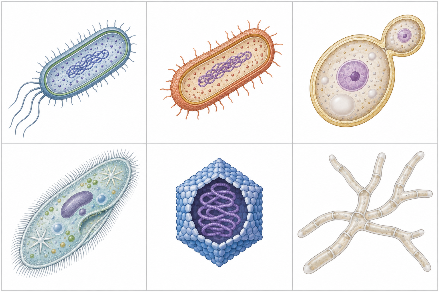
Choose an image and connect its structure to the explanation. AI-generated schematics with no common scale; these are not micrographs or diagnostic images.
Bacterial cell
Notice the DNA region without a membrane-bound nucleus. This is one schematic rod, not the shape of every bacterium. Bacteria may also be round or curved; the picture does not identify a species or prove disease.
Original image and limitations
Sizes, colours and details are simplified for conceptual comparison. The archaeal outline is not an identification criterion. Labels and explanations use your selected language.
Read each row for cell organisation and typical replication. These are selected teaching groups, not an exhaustive taxonomy. Compare the PNG structures with the statements, then select a numbered explanation.
New to diagrams? Read this first
Look at the whole drawing once without clicking anything. Ask yourself only: what kind of picture is this (a comparison, a route, a sequence, a table)?
Press Play, or use the arrows, to move through the numbered parts in order. The drawing stays still; only the highlight moves.
Click a number, or its name in the list below, to read what that part is and why it matters. Use “Hide labels” when you want to test your memory.
More controls
01 / 5Bacteria: organisation and growth
If the drawing is wider than this panel, scroll it sideways. Labels stay readable; the numbered explanations below show the same ideas.
Bacteria: organisation and growth
A bacterium has a membrane, cytoplasm, ribosomes and genetic material, but no membrane-bound nucleus. Most bacteria also have a peptidoglycan cell wall; exceptions such as Mycoplasma lack that wall.
Most multiply by binary fission after copying their DNA. Growth needs suitable nutrients and conditions; some bacteria require living host cells. Bacteria occur in soil, water and the body, and can be carried in air. Many recycle nutrients or form part of the microbiota; some cause infection. A colony on a plate is visible growth from many cells, not one enlarged bacterium.
Conceptual comparison, not to scale. The five selected groups are not equal taxonomic ranks or the whole field. Bacteria and archaea can look similar; identity needs appropriate tests. Most individual viruses cannot be resolved by an ordinary optical microscope.
Key points to remember
✓This section compares bacteria, archaea, fungi, protozoa and viruses; these five groups are not an exhaustive classification.
✓Bacteria are prokaryotic cells; most multiply by binary fission when their requirements for growth are met.
✓Archaea are prokaryotes but a separate domain, defined by a unique ribosomal RNA sequence and by different membrane and wall chemistry; methanogens make methane.
✓Fungi are eukaryotes: yeasts may be single cells, whereas moulds form hyphae and mycelium.
✓Protozoa are diverse unicellular eukaryotes; viruses are acellular and replicate using host cells.
Go deeper
Worked example
Sorting an unknown organism into one of the five groups. A teaching laboratory compares three samples: a broth with small cells, a plate with branching fungal filaments, and a respiratory sample with no growth on routine bacterial agar. Use the observations to suggest the next investigation without pretending they identify every agent.
Describe what is actually observed. Broth turbidity and colonies suggest growth, but microscopic examination is needed to describe cells. No growth in the respiratory sample is inconclusive: antibiotics, sampling and unsuitable conditions can all reduce recovery.
Use supported structural evidence. Branching hyphae support a mould identification. Confirmed absence of a membrane-bound nucleus suggests a prokaryote, but bacterial and archaeal cells cannot be separated just by their outline.
Choose a discriminating test. Ribosomal RNA sequence and membrane chemistry can support domain identification. An extreme habitat is not enough: bacteria also tolerate extremes, and some bacteria lack a wall. A suspected virus needs a suitable specific test, not a diagnosis from no growth alone.
Keep identification separate from treatment. Group information guides further testing; it does not prove that the organism causes disease or that a particular drug will work. Clinical assessment and, when appropriate, susceptibility results guide the prescriber.
The group is not an end in itself. It is the first fork in a decision tree: cell or no cell, prokaryote or eukaryote, and then which structure a drug can attack. That is why section 1.2 comes before the sections on the bacterial cell and on viruses.
Common misconception
A virus is not a small bacterium, an archaeon is not simply a bacterium from a hot spring, and yeast does not need hyphae to be a fungus. Failed routine culture does not prove a viral infection. Use cell organisation and specific tests, and distinguish the five groups selected here from the wider field of microbiology.
Check your reasoning
Think of one infection you have seen or read about. Which of the five groups causes it, and what does that group tell you about which medicine could work and which could not?
Reveal an answer
Influenza is viral: ordinary antibacterial drugs do not target its replication. A bacterial wound infection is different, but the presence of bacterial structures still does not guarantee that a chosen antibiotic will work. Susceptibility, infection site and the patient’s condition matter. A group is a guide to investigation, not a complete treatment decision.
Where this comes from
BMS 114 General Microbiology I lecture notes (supplied) · “The major groups of microorganisms”, lines 225–278
Correction to the supplied notes — the number of groups · The supplied notes announce six groups but name five. This lesson compares the named five while explicitly retaining algae and helminths within wider microbiology; the list is not an exhaustive taxonomy.
Correction in emphasis — archaea are a separate domain · Correction to the supplied notes: line 250 calls archaea phylogenetically related prokaryotes and stops there. Woese’s comparison of 16S ribosomal RNA places them in a separate domain, with different membrane lipids and without peptidoglycan; the notes understate the difference.
Beyond the supplied notes — drug targets and survival outside the host · selective toxicity and the targets of beta-lactams, aminoglycosides, tetracyclines, macrolides, sulphonamides, trimethoprim, fluoroquinolones, azoles, amphotericin B, echinocandins and antivirals; the three-domain system (Woese, 1977) and Whittaker’s five kingdoms (1969); endospores, protozoal cysts and enveloped versus non-enveloped viruses. The notes at lines 225–278 name the groups, their cell type, their multiplication and their habitats, but do not explain these mechanisms.
Lesson progress is saved on this device for the current learner. Practice answers stay separate and are not stored.
About this course and its sources
Bilingual lessons organised from the supplied BMS 114 General Microbiology I lecture notes (Mr Ngotelo F. Ambene, Mr Lyonga B. Lyonga & Mr Fumbui Louis Ngong). Diagrams are original educational schematics; sections that correct the notes or go beyond them say so in their sources. Nursing and French-language review are pending.